7S5E | pdb_00007s5e

Crystal structure of a guanylate kinase from Stenotrophomonas maltophilia K279a with heterogeneous ligand states of GMP/ADP, GMP/-, GDP/-, and GMP/ATPgS


Experimental Data Snapshot

  • Method: X-RAY DIFFRACTION
  • Resolution: 2.40 Å
  • R-Value Free: 
    0.265 (Depositor), 0.264 (DCC) 
  • R-Value Work: 
    0.226 (Depositor), 0.226 (DCC) 
  • R-Value Observed: 
    0.227 (Depositor) 

Starting Model: experimental
View more details

wwPDB Validation 3D Report Full Report

Validation slider image for 7S5E

Ligand Structure Quality Assessment 


This is version 1.1 of the entry. See complete history

Literature

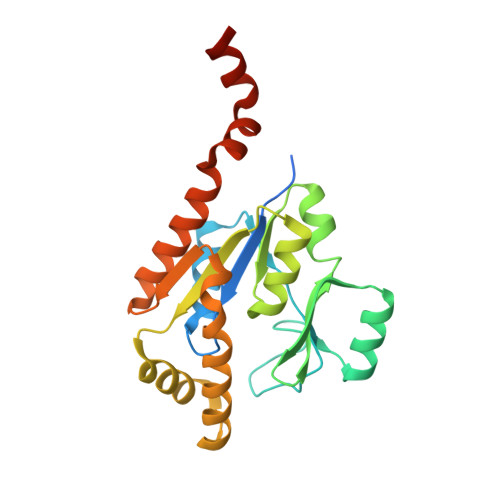
Crystal structure of a guanylate kinase from Stenotrophomonas maltophilia K279c with heterogeneous ligand states of GMP/ADP, GMP/-, GDP/-, and GMP/ATPgS.

Edwards, T.E.Horanyi, P.S.Abendroth, J.Lorimer, D.D.Seattle Structural Genomics Center for Infectious Disease (SSGCID)

To be published.

Macromolecule Content 

  • Total Structure Weight: 206.67 kDa 
  • Atom Count: 12,787 
  • Modeled Residue Count: 1,612 
  • Deposited Residue Count: 1,832 
  • Unique protein chains: 1

Macromolecules

Find similar proteins by:|  3D Structure
Entity ID: 1
MoleculeChains  Sequence LengthOrganismDetailsImage
Guanylate kinase
A, B, C, D, E
A, B, C, D, E, F, G, H
229Stenotrophomonas maltophilia K279aMutation(s): 0 
Gene Names: gmkSmlt3842
EC: 2.7.4.8
UniProt
Find proteins for B2FT06 (Stenotrophomonas maltophilia (strain K279a))
Explore B2FT06 
Go to UniProtKB:  B2FT06
Entity Groups
Sequence Clusters30% Identity50% Identity70% Identity90% Identity95% Identity100% Identity
UniProt GroupB2FT06
Sequence Annotations
Expand
Reference Sequence

Small Molecules

Ligands 4 Unique
IDChains Name / Formula / InChI Key2D Diagram3D Interactions
AGS
(Subject of Investigation/LOI)

Query on AGS


Download:Ideal Coordinates CCD File
N [auth D],
T [auth H]
PHOSPHOTHIOPHOSPHORIC ACID-ADENYLATE ESTER
C10 H16 N5 O12 P3 S
NLTUCYMLOPLUHL-KQYNXXCUSA-N
GDP
(Subject of Investigation/LOI)

Query on GDP


Download:Ideal Coordinates CCD File
L [auth C],
R [auth G]
GUANOSINE-5'-DIPHOSPHATE
C10 H15 N5 O11 P2
QGWNDRXFNXRZMB-UUOKFMHZSA-N
ADP
(Subject of Investigation/LOI)

Query on ADP


Download:Ideal Coordinates CCD File
J [auth A],
P [auth E]
ADENOSINE-5'-DIPHOSPHATE
C10 H15 N5 O10 P2
XTWYTFMLZFPYCI-KQYNXXCUSA-N
5GP
(Subject of Investigation/LOI)

Query on 5GP


Download:Ideal Coordinates CCD File
I [auth A]
K [auth B]
M [auth D]
O [auth E]
Q [auth F]
I [auth A],
K [auth B],
M [auth D],
O [auth E],
Q [auth F],
S [auth H]
GUANOSINE-5'-MONOPHOSPHATE
C10 H14 N5 O8 P
RQFCJASXJCIDSX-UUOKFMHZSA-N

Experimental Data & Validation

Experimental Data

  • Method: X-RAY DIFFRACTION
  • Resolution: 2.40 Å
  • R-Value Free:  0.265 (Depositor), 0.264 (DCC) 
  • R-Value Work:  0.226 (Depositor), 0.226 (DCC) 
  • R-Value Observed: 0.227 (Depositor) 
Space Group: P 1 21 1
Unit Cell:
Length ( Å )Angle ( ˚ )
a = 97.16α = 90
b = 100.53β = 95.21
c = 92.25γ = 90
Software Package:
Software NamePurpose
XDSdata reduction
XSCALEdata scaling
MOLREPphasing
PHENIXrefinement
PDB_EXTRACTdata extraction

Structure Validation

View Full Validation Report


Ligand Structure Quality Assessment 


Entry History 

& Funding Information

Deposition Data


Funding OrganizationLocationGrant Number
National Institutes of Health/National Institute Of Allergy and Infectious Diseases (NIH/NIAID)United States--

Revision History  (Full details and data files)

  • Version 1.0: 2021-09-22
    Type: Initial release
  • Version 1.1: 2023-10-18
    Changes: Data collection, Refinement description