7S4W

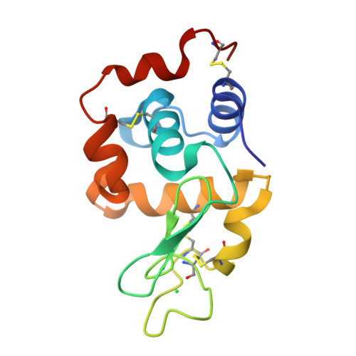
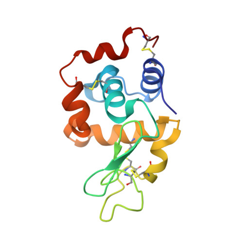
Serial Macromolecular Crystallography at ALBA Synchrotron Light Source - Lysozyme


Experimental Data Snapshot

  • Method: X-RAY DIFFRACTION
  • Resolution: 2.10 Å
  • R-Value Free: 0.232 
  • R-Value Work: 0.178 
  • R-Value Observed: 0.184 

Starting Model: experimental
View more details

wwPDB Validation   3D Report Full Report


This is version 1.3 of the entry. See complete history


Literature

Serial macromolecular crystallography at ALBA Synchrotron Light Source.

Martin-Garcia, J.M.Botha, S.Hu, H.Jernigan, R.Castellvi, A.Lisova, S.Gil, F.Calisto, B.Crespo, I.Roy-Chowdhury, S.Grieco, A.Ketawala, G.Weierstall, U.Spence, J.Fromme, P.Zatsepin, N.Boer, D.R.Carpena, X.

(2022) J Synchrotron Radiat 29: 896-907

  • DOI: https://doi.org/10.1107/S1600577522002508
  • Primary Citation of Related Structures:  
    7S4R, 7S4W, 7S4Y, 7S4Z, 7S50

  • PubMed Abstract: 

    The increase in successful adaptations of serial crystallography at synchrotron radiation sources continues. To date, the number of serial synchrotron crystallography (SSX) experiments has grown exponentially, with over 40 experiments reported so far. In this work, we report the first SSX experiments with viscous jets conducted at ALBA beamline BL13-XALOC. Small crystals (15-30 µm) of five soluble proteins (lysozyme, proteinase K, phycocyanin, insulin and α-spectrin-SH3 domain) were suspended in lipidic cubic phase (LCP) and delivered to the X-ray beam with a high-viscosity injector developed at Arizona State University. Complete data sets were collected from all proteins and their high-resolution structures determined. The high quality of the diffraction data collected from all five samples, and the lack of specific radiation damage in the structures obtained in this study, confirm that the current capabilities at the beamline enables atomic resolution determination of protein structures from microcrystals as small as 15 µm using viscous jets at room temperature. Thus, BL13-XALOC can provide a feasible alternative to X-ray free-electron lasers when determining snapshots of macromolecular structures.


  • Organizational Affiliation

    Center for Applied Structural Discovery, Biodesign Institute, Arizona State University, Tempe, AZ, USA.


Macromolecules
Find similar proteins by:  (by identity cutoff)  |  3D Structure
Entity ID: 1
MoleculeChains Sequence LengthOrganismDetailsImage
Lysozyme C129Gallus gallusMutation(s): 0 
Gene Names: LYZ
EC: 3.2.1.17
UniProt
Find proteins for P00698 (Gallus gallus)
Explore P00698 
Go to UniProtKB:  P00698
Entity Groups  
Sequence Clusters30% Identity50% Identity70% Identity90% Identity95% Identity100% Identity
UniProt GroupP00698
Sequence Annotations
Expand
  • Reference Sequence
Small Molecules
Ligands 1 Unique
IDChains Name / Formula / InChI Key2D Diagram3D Interactions
CL
Query on CL

Download Ideal Coordinates CCD File 
B [auth A]CHLORIDE ION
Cl
VEXZGXHMUGYJMC-UHFFFAOYSA-M
Experimental Data & Validation

Experimental Data

  • Method: X-RAY DIFFRACTION
  • Resolution: 2.10 Å
  • R-Value Free: 0.232 
  • R-Value Work: 0.178 
  • R-Value Observed: 0.184 
  • Space Group: P 43 21 2
Unit Cell:
Length ( Å )Angle ( ˚ )
a = 79α = 90
b = 79β = 90
c = 38.2γ = 90
Software Package:
Software NamePurpose
REFMACrefinement
CrystFELdata reduction
Aimlessdata scaling
MOLREPphasing

Structure Validation

View Full Validation Report


Entry History & Funding Information

Deposition Data


Funding OrganizationLocationGrant Number
National Science Foundation (NSF, United States)1231306
Other government2019-T1BMD-15552

Revision History  (Full details and data files)

  • Version 1.0: 2021-10-27
    Type: Initial release
  • Version 1.1: 2022-05-18
    Changes: Database references
  • Version 1.2: 2023-10-18
    Changes: Data collection, Refinement description
  • Version 1.3: 2024-11-06
    Changes: Structure summary