7S49 | pdb_00007s49

Crystal Structure of Inhibitor-bound Galactokinase

  • Classification: Transferase/Inhibitor
  • Organism(s): Homo sapiens
  • Expression System: Escherichia coli
  • Mutation(s): Yes 

  • Deposited: 2021-09-08 Released: 2021-09-29 
  • Deposition Author(s): Whitby, F.G.
  • Funding Organization(s): National Institutes of Health/National Institute of General Medical Sciences (NIH/NIGMS)

Experimental Data Snapshot

  • Method: X-RAY DIFFRACTION
  • Resolution: 2.20 Å
  • R-Value Free: 
    0.226 (Depositor), 0.226 (DCC) 
  • R-Value Work: 
    0.158 (Depositor), 0.158 (DCC) 
  • R-Value Observed: 
    0.160 (Depositor) 

Starting Model: experimental
View more details

wwPDB Validation 3D Report Full Report

Validation slider image for 7S49

Ligand Structure Quality Assessment 


This is version 1.4 of the entry. See complete history

Literature

Structure-Based Optimization of Small Molecule Human Galactokinase Inhibitors.

Liu, L.Tang, M.Pragani, R.Whitby, F.G.Zhang, Y.Q.Balakrishnan, B.Fang, Y.Karavadhi, S.Tao, D.LeClair, C.A.Hall, M.D.Marugan, J.J.Boxer, M.Shen, M.Hill, C.P.Lai, K.Patnaik, S.

(2021) J Med Chem 64: 13551-13571

  • DOI: https://doi.org/10.1021/acs.jmedchem.1c00945
  • Primary Citation Related Structures: 
    7RCL, 7RCM, 7S49, 7S4C

  • PubMed Abstract: 

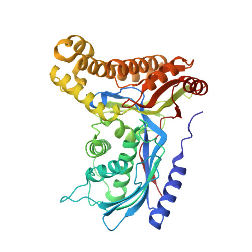
    Classic galactosemia is a rare disease caused by inherited deficiency of galactose-1 phosphate uridylyltransferase (GALT). Accumulation of galactose-1 phosphate (gal-1P) is thought to be the major cause of the chronic complications associated with this disease, which currently has no treatment. Inhibiting galactokinase (GALK1), the enzyme that generates galactose-1 phosphate, has been proposed as a novel strategy for treating classic galactosemia. Our previous work identified a highly selective unique dihydropyrimidine inhibitor against GALK1. With the determination of a co-crystal structure of this inhibitor with human GALK1, we initiated a structure-based structure-activity relationship (SAR) optimization campaign that yielded novel analogs with potent biochemical inhibition (IC 50 < 100 nM). Lead compounds were also able to prevent gal-1P accumulation in patient-derived cells at low micromolar concentrations and have pharmacokinetic properties suitable for evaluation in rodent models of galactosemia.


  • Organizational Affiliation
    • National Center for Advancing Translational Sciences, National Institutes of Health, 9800 Medical Center Drive, Rockville, Maryland 20850, United States.

Macromolecule Content 

  • Total Structure Weight: 85.96 kDa 
  • Atom Count: 6,267 
  • Modeled Residue Count: 782 
  • Deposited Residue Count: 784 
  • Unique protein chains: 1

Macromolecules

Find similar proteins by:|  3D Structure
Entity ID: 1
MoleculeChains  Sequence LengthOrganismDetailsImage
Galactokinase
A, B
392Homo sapiensMutation(s): 2 
Gene Names: GALK1GALK
EC: 2.7.1.6
UniProt & NIH Common Fund Data Resources
Find proteins for P51570 (Homo sapiens)
Explore P51570 
Go to UniProtKB:  P51570
PHAROS:  P51570
GTEx:  ENSG00000108479 
Entity Groups
Sequence Clusters30% Identity50% Identity70% Identity90% Identity95% Identity100% Identity
UniProt GroupP51570
Sequence Annotations
Expand
Reference Sequence

Small Molecules

Ligands 4 Unique
IDChains Name / Formula / InChI Key2D Diagram3D Interactions
4QI
(Subject of Investigation/LOI)

Query on 4QI


Download:Ideal Coordinates CCD File
D [auth A],
J [auth B]
(4R)-2-[(1,3-benzoxazol-2-yl)amino]-4-(4-chloro-1H-pyrazol-5-yl)-4,6,7,8-tetrahydroquinazolin-5(1H)-one
C18 H15 Cl N6 O2
ORBGGGFENULXFO-MRXNPFEDSA-N
GLA

Query on GLA


Download:Ideal Coordinates CCD File
C [auth A],
I [auth B]
alpha-D-galactopyranose
C6 H12 O6
WQZGKKKJIJFFOK-PHYPRBDBSA-N
PO4

Query on PO4


Download:Ideal Coordinates CCD File
E [auth A],
F [auth A],
G [auth A],
K [auth B]
PHOSPHATE ION
O4 P
NBIIXXVUZAFLBC-UHFFFAOYSA-K
NA

Query on NA


Download:Ideal Coordinates CCD File
H [auth A],
L [auth B]
SODIUM ION
Na
FKNQFGJONOIPTF-UHFFFAOYSA-N

Experimental Data & Validation

Experimental Data

  • Method: X-RAY DIFFRACTION
  • Resolution: 2.20 Å
  • R-Value Free:  0.226 (Depositor), 0.226 (DCC) 
  • R-Value Work:  0.158 (Depositor), 0.158 (DCC) 
  • R-Value Observed: 0.160 (Depositor) 
Space Group: P 61 2 2
Unit Cell:
Length ( Å )Angle ( ˚ )
a = 129.407α = 90
b = 129.407β = 90
c = 240.548γ = 120
Software Package:
Software NamePurpose
DENZOdata reduction
SCALEPACKdata scaling
REFMACrefinement
PDB_EXTRACTdata extraction
PHENIXphasing

Structure Validation

View Full Validation Report


Ligand Structure Quality Assessment 


Entry History 

& Funding Information

Deposition Data

  • Released Date: 2021-09-29 
  • Deposition Author(s): Whitby, F.G.

Funding OrganizationLocationGrant Number
National Institutes of Health/National Institute of General Medical Sciences (NIH/NIGMS)United StatesRO1HD074844

Revision History  (Full details and data files)

  • Version 1.0: 2021-09-29
    Type: Initial release
  • Version 1.1: 2021-10-06
    Changes: Database references
  • Version 1.2: 2023-02-01
    Changes: Advisory, Database references
  • Version 1.3: 2023-10-25
    Changes: Data collection, Refinement description
  • Version 1.4: 2024-10-30
    Changes: Structure summary