7S26 | pdb_00007s26

ROCK1 IN COMPLEX WITH LIGAND G5018


Experimental Data Snapshot

  • Method: X-RAY DIFFRACTION
  • Resolution: 2.74 Å
  • R-Value Free: 
    0.269 (Depositor), 0.269 (DCC) 
  • R-Value Work: 
    0.247 (Depositor), 0.239 (DCC) 
  • R-Value Observed: 
    0.247 (Depositor) 

wwPDB Validation 3D Report Full Report

Validation slider image for 7S26

Ligand Structure Quality Assessment 


This is version 1.2 of the entry. See complete history

Literature

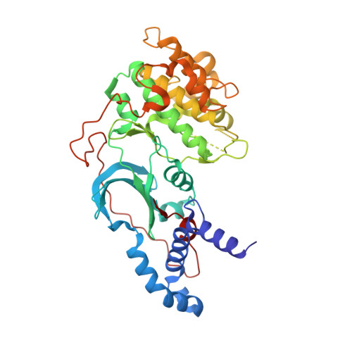
Chemical space docking enables large-scale structure-based virtual screening to discover ROCK1 kinase inhibitors.

Beroza, P.Crawford, J.J.Ganichkin, O.Gendelev, L.Harris, S.F.Klein, R.Miu, A.Steinbacher, S.Klingler, F.M.Lemmen, C.

(2022) Nat Commun 13: 6447-6447

  • DOI: https://doi.org/10.1038/s41467-022-33981-8
  • Primary Citation Related Structures: 
    7S25, 7S26

  • PubMed Abstract: 

    With the ever-increasing number of synthesis-on-demand compounds for drug lead discovery, there is a great need for efficient search technologies. We present the successful application of a virtual screening method that combines two advances: (1) it avoids full library enumeration (2) products are evaluated by molecular docking, leveraging protein structural information. Crucially, these advances enable a structure-based technique that can efficiently explore libraries with billions of molecules and beyond. We apply this method to identify inhibitors of ROCK1 from almost one billion commercially available compounds. Out of 69 purchased compounds, 27 (39%) have K i values < 10 µM. X-ray structures of two leads confirm their docked poses. This approach to docking scales roughly with the number of reagents that span a chemical space and is therefore multiple orders of magnitude faster than traditional docking.


  • Organizational Affiliation
    • Discovery Chemistry, Genentech, South San Francisco, USA. berozap@gene.com.

Macromolecule Content 

  • Total Structure Weight: 185.55 kDa 
  • Atom Count: 11,460 
  • Modeled Residue Count: 1,379 
  • Deposited Residue Count: 1,592 
  • Unique protein chains: 1

Macromolecules

Find similar proteins by:|  3D Structure
Entity ID: 1
MoleculeChains  Sequence LengthOrganismDetailsImage
Rho-associated protein kinase 1
A, B, C, D
398Homo sapiensMutation(s): 0 
Gene Names: ROCK1
EC: 2.7.11.1 (PDB Primary Data), 2.7.11.39 (UniProt)
UniProt & NIH Common Fund Data Resources
Find proteins for Q13464 (Homo sapiens)
Explore Q13464 
Go to UniProtKB:  Q13464
PHAROS:  Q13464
GTEx:  ENSG00000067900 
Entity Groups
Sequence Clusters30% Identity50% Identity70% Identity90% Identity95% Identity100% Identity
UniProt GroupQ13464
Sequence Annotations
Expand
Reference Sequence

Experimental Data & Validation

Experimental Data

  • Method: X-RAY DIFFRACTION
  • Resolution: 2.74 Å
  • R-Value Free:  0.269 (Depositor), 0.269 (DCC) 
  • R-Value Work:  0.247 (Depositor), 0.239 (DCC) 
  • R-Value Observed: 0.247 (Depositor) 
Space Group: P 1 21 1
Unit Cell:
Length ( Å )Angle ( ˚ )
a = 57.141α = 90
b = 101.596β = 91.98
c = 182.663γ = 90
Software Package:
Software NamePurpose
BUSTERrefinement
Aimlessdata scaling
PDB_EXTRACTdata extraction
XDSdata reduction
MOLREPphasing

Structure Validation

View Full Validation Report


Ligand Structure Quality Assessment 


Entry History 

& Funding Information

Deposition Data


Funding OrganizationLocationGrant Number
Not funded--

Revision History  (Full details and data files)

  • Version 1.0: 2022-10-05
    Type: Initial release
  • Version 1.1: 2023-04-19
    Changes: Database references
  • Version 1.2: 2024-05-22
    Changes: Data collection