7RY5 | pdb_00007ry5

Cellular Retinoic Acid Binding Protein II with Bound Inhibitor 4-[6-({4-[(fluorosulfonyl)oxy]phenyl}ethynyl)-4,4-dimethyl-3,4-dihydroquinolin-1(2H)-yl]-4-oxobutanoic acid


Experimental Data Snapshot

  • Method: X-RAY DIFFRACTION
  • Resolution: 2.00 Å
  • R-Value Free: 
    0.233 (Depositor), 0.243 (DCC) 
  • R-Value Work: 
    0.175 (Depositor), 0.185 (DCC) 

Starting Model: experimental
View more details

wwPDB Validation 3D Report Full Report

Validation slider image for 7RY5

Ligand Structure Quality Assessment 


This is version 1.3 of the entry. See complete history

Literature

Development of an Enzyme-Inhibitor Reaction Using Cellular Retinoic Acid Binding Protein II for One-Pot Megamolecule Assembly.

Kimmel, B.R.Mrksich, M.

(2021) Chemistry 27: 17843-17848

  • DOI: https://doi.org/10.1002/chem.202103059
  • Primary Citation Related Structures: 
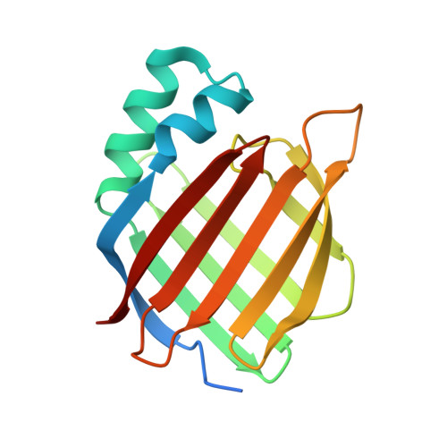
    7RY5

  • PubMed Abstract: 

    This paper presents an enzyme building block for the assembly of megamolecules. The system is based on the inhibition of the human-derived cellular retinoic acid binding protein II (CRABP2) domain. We synthesized a synthetic retinoid bearing an arylfluorosulfate group, which uses sulfur fluoride exchange click chemistry to covalently inhibit CRABP2. We conjugated both the inhibitor and a fluorescein tag to an oligo(ethylene glycol) backbone and measured a second-order rate constant for the protein inhibition reaction of approximately 3,600 M -1 s -1 . We used this new enzyme-inhibitor pair to assemble multi-protein structures in one-pot reactions using three orthogonal assembly chemistries to demonstrate exact control over the placement of protein domains within a single, homogeneous molecule. This work enables a new dimension of control over specificity, orientation, and stoichiometry of protein domains within atomically precise nanostructures.


  • Organizational Affiliation
    • Department of Chemical and Biological Engineering, Northwestern University, 2145 Sheridan Road, Evanston, IL, 60208, USA.

Macromolecule Content 

  • Total Structure Weight: 35.71 kDa 
  • Atom Count: 2,550 
  • Modeled Residue Count: 281 
  • Deposited Residue Count: 302 
  • Unique protein chains: 1

Macromolecules

Find similar proteins by:|  3D Structure
Entity ID: 1
MoleculeChains  Sequence LengthOrganismDetailsImage
Cellular retinoic acid-binding protein 2A [auth AAA],
B [auth BBB]
151Homo sapiensMutation(s): 0 
Gene Names: CRABP2
UniProt & NIH Common Fund Data Resources
Find proteins for P29373 (Homo sapiens)
Explore P29373 
Go to UniProtKB:  P29373
PHAROS:  P29373
GTEx:  ENSG00000143320 
Entity Groups
Sequence Clusters30% Identity50% Identity70% Identity90% Identity95% Identity100% Identity
UniProt GroupP29373
Sequence Annotations
Expand
Reference Sequence

Small Molecules

Ligands 1 Unique
IDChains Name / Formula / InChI Key2D Diagram3D Interactions
80V
(Subject of Investigation/LOI)

Query on 80V


Download:Ideal Coordinates CCD File
C [auth AAA],
D [auth BBB]
4-[6-({4-[(fluorosulfonyl)oxy]phenyl}ethynyl)-4,4-dimethyl-3,4-dihydroquinolin-1(2H)-yl]-4-oxobutanoic acid
C23 H22 F N O6 S
OJLXEIKKMUTWLF-UHFFFAOYSA-N

Experimental Data & Validation

Experimental Data

  • Method: X-RAY DIFFRACTION
  • Resolution: 2.00 Å
  • R-Value Free:  0.233 (Depositor), 0.243 (DCC) 
  • R-Value Work:  0.175 (Depositor), 0.185 (DCC) 
Space Group: P 2 21 21
Unit Cell:
Length ( Å )Angle ( ˚ )
a = 36.604α = 90
b = 69.655β = 90
c = 120.845γ = 90
Software Package:
Software NamePurpose
REFMACrefinement
HKL-2000data reduction
HKL-2000data scaling
PHASERphasing

Structure Validation

View Full Validation Report


Ligand Structure Quality Assessment 


Entry History 

& Funding Information

Deposition Data


Funding OrganizationLocationGrant Number
National Science Foundation (NSF, United States)United StatesDGE-1842165

Revision History  (Full details and data files)

  • Version 1.0: 2021-12-15
    Type: Initial release
  • Version 1.1: 2022-01-05
    Changes: Database references
  • Version 1.2: 2023-10-18
    Changes: Data collection, Refinement description
  • Version 1.3: 2024-11-06
    Changes: Structure summary