7RUG | pdb_00007rug

Human SERINC3-DeltaICL4


Experimental Data Snapshot

  • Method: ELECTRON MICROSCOPY
  • Resolution: 4.70 Å
  • Aggregation State: PARTICLE 
  • Reconstruction Method: SINGLE PARTICLE 

wwPDB Validation 3D Report Full Report

Validation slider image for 7RUG

This is version 1.1 of the entry. See complete history

Literature

Antiviral HIV-1 SERINC restriction factors disrupt virus membrane asymmetry.

Leonhardt, S.A.Purdy, M.D.Grover, J.R.Yang, Z.Poulos, S.McIntire, W.E.Tatham, E.A.Erramilli, S.K.Nosol, K.Lai, K.K.Ding, S.Lu, M.Uchil, P.D.Finzi, A.Rein, A.Kossiakoff, A.A.Mothes, W.Yeager, M.

(2023) Nat Commun 14: 4368-4368

  • DOI: https://doi.org/10.1038/s41467-023-39262-2
  • Primary Citation Related Structures: 
    7RU6, 7RUG

  • PubMed Abstract: 

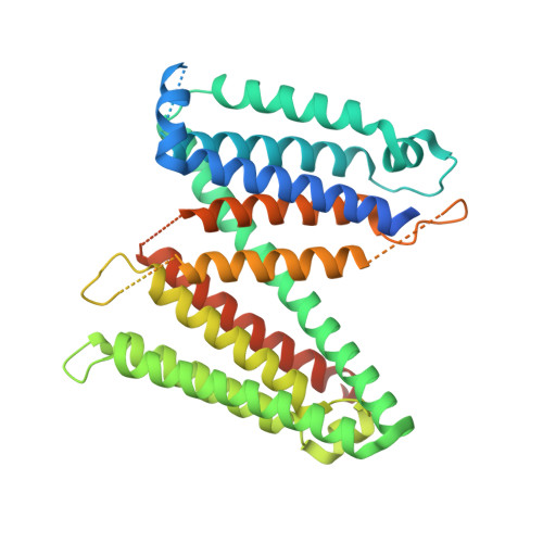
    The host proteins SERINC3 and SERINC5 are HIV-1 restriction factors that reduce infectivity when incorporated into the viral envelope. The HIV-1 accessory protein Nef abrogates incorporation of SERINCs via binding to intracellular loop 4 (ICL4). Here, we determine cryoEM maps of full-length human SERINC3 and an ICL4 deletion construct, which reveal that hSERINC3 is comprised of two α-helical bundles connected by a ~ 40-residue, highly tilted, "crossmember" helix. The design resembles non-ATP-dependent lipid transporters. Consistently, purified hSERINCs reconstituted into proteoliposomes induce flipping of phosphatidylserine (PS), phosphatidylethanolamine and phosphatidylcholine. Furthermore, SERINC3, SERINC5 and the scramblase TMEM16F expose PS on the surface of HIV-1 and reduce infectivity, with similar results in MLV. SERINC effects in HIV-1 and MLV are counteracted by Nef and GlycoGag, respectively. Our results demonstrate that SERINCs are membrane transporters that flip lipids, resulting in a loss of membrane asymmetry that is strongly correlated with changes in Env conformation and loss of infectivity.


  • Organizational Affiliation
    • The Phillip and Patricia Frost Institute for Chemistry and Molecular Science, University of Miami, Coral Gables, FL, 33146, USA.

Macromolecule Content 

  • Total Structure Weight: 78.1 kDa 
  • Atom Count: 2,701 
  • Modeled Residue Count: 331 
  • Deposited Residue Count: 709 
  • Unique protein chains: 2

Macromolecules

Find similar proteins by:|  3D Structure
Entity ID: 1
MoleculeChains  Sequence LengthOrganismDetailsImage
Serine incorporator 3468Homo sapiensMutation(s): 0 
Gene Names: SERINC3DIFF33TDE1SBBI99
Membrane Entity: Yes 
UniProt & NIH Common Fund Data Resources
Find proteins for Q13530 (Homo sapiens)
Explore Q13530 
Go to UniProtKB:  Q13530
PHAROS:  Q13530
GTEx:  ENSG00000132824 
Entity Groups
Sequence Clusters30% Identity50% Identity70% Identity90% Identity95% Identity100% Identity
UniProt GroupQ13530
Sequence Annotations
Expand
Reference Sequence
Find similar proteins by:|  3D Structure
Entity ID: 2
MoleculeChains  Sequence LengthOrganismDetailsImage
SiAB [auth L]241synthetic constructMutation(s): 0 

Experimental Data & Validation

Experimental Data

  • Method: ELECTRON MICROSCOPY
  • Resolution: 4.70 Å
  • Aggregation State: PARTICLE 
  • Reconstruction Method: SINGLE PARTICLE 
EM Software:
TaskSoftware PackageVersion
RECONSTRUCTIONRELION3.1
MODEL REFINEMENTPHENIX1.20.1-4487

Structure Validation

View Full Validation Report


Entry History 

& Funding Information

Deposition Data


Funding OrganizationLocationGrant Number
National Institutes of Health/National Institute Of Allergy and Infectious Diseases (NIH/NIAID)United States5P50AI150464-15

Revision History  (Full details and data files)

  • Version 1.0: 2022-08-24
    Type: Initial release
  • Version 1.1: 2023-09-06
    Changes: Data collection, Database references