7RR3 | pdb_00007rr3

Structure of Deep-Sea Phage NrS-1 Primase-Polymerase N300 in complex with calcium and ddCTP


Experimental Data Snapshot

  • Method: X-RAY DIFFRACTION
  • Resolution: 2.24 Å
  • R-Value Free: 
    0.287 (Depositor), 0.287 (DCC) 
  • R-Value Work: 
    0.235 (Depositor), 0.235 (DCC) 
  • R-Value Observed: 
    0.238 (Depositor) 

wwPDB Validation 3D Report Full Report

Validation slider image for 7RR3

Ligand Structure Quality Assessment 


This is version 1.2 of the entry. See complete history

Literature

Molecular Dissection of the Primase and Polymerase Activities of Deep-Sea Phage NrS-1 Primase-Polymerase.

Huang, F.Lu, X.Yu, C.Sliz, P.Wang, L.Zhu, B.

(2021) Front Microbiol 12: 766612-766612

  • DOI: https://doi.org/10.3389/fmicb.2021.766612
  • Primary Citation Related Structures: 
    7RR3

  • PubMed Abstract: 

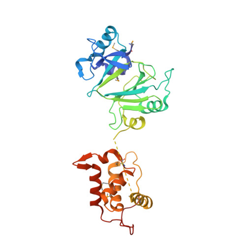
    PrimPols are a class of primases that belong to the archaeo-eukaryotic primase (AEP) superfamily but have both primase and DNA polymerase activities. Replicative polymerase from NrS-1 phage (NrSPol) is a representative of the PrimPols. In this study, we identified key residues for the catalytic activity of NrSPol and found that a loop in NrSPol functionally replaces the zinc finger motif that is commonly found in other AEP family proteins. A helix bundle domain (HBD), conserved in the AEP superfamily, was recently reported to bind to the primase recognition site and to be crucial for initiation of primer synthesis. We found that NrSPol can recognize different primase recognition sites, and that the initiation site for primer synthesis is not stringent, suggesting that the HBD conformation is flexible. More importantly, we found that although the HBD-inactivating mutation impairs the primase activity of NrSPol, it significantly enhances the DNA polymerase activity, indicating that the HBD hinders the DNA polymerase activity. The conflict between the primase activity and the DNA polymerase activity in a single protein with the same catalytic domain may be one reason for why DNA polymerases are generally unable to synthesize DNA de novo .


  • Organizational Affiliation
    • Key Laboratory of Molecular Biophysics, The Ministry of Education, College of Life Science and Technology and Shenzhen College, Huazhong University of Science and Technology, Wuhan, China.

Macromolecule Content 

  • Total Structure Weight: 33.74 kDa 
  • Atom Count: 2,190 
  • Modeled Residue Count: 275 
  • Deposited Residue Count: 289 
  • Unique protein chains: 1

Macromolecules

Find similar proteins by:|  3D Structure
Entity ID: 1
MoleculeChains  Sequence LengthOrganismDetailsImage
Primase289Nitratiruptor phage NrS-1Mutation(s): 0 
EC: 2.7.7.7 (UniProt), 3.6.4.12 (UniProt), 2.7.7 (UniProt)
UniProt
Find proteins for M5AAG8 (Nitratiruptor phage NrS-1)
Explore M5AAG8 
Go to UniProtKB:  M5AAG8
Entity Groups
Sequence Clusters30% Identity50% Identity70% Identity90% Identity95% Identity100% Identity
UniProt GroupM5AAG8
Sequence Annotations
Expand
Reference Sequence

Experimental Data & Validation

Experimental Data

  • Method: X-RAY DIFFRACTION
  • Resolution: 2.24 Å
  • R-Value Free:  0.287 (Depositor), 0.287 (DCC) 
  • R-Value Work:  0.235 (Depositor), 0.235 (DCC) 
  • R-Value Observed: 0.238 (Depositor) 
Space Group: P 21 21 21
Unit Cell:
Length ( Å )Angle ( ˚ )
a = 56.452α = 90
b = 71.58β = 90
c = 72.962γ = 90
Software Package:
Software NamePurpose
PHENIXrefinement
xia2data reduction
xia2data scaling
PHENIXphasing

Structure Validation

View Full Validation Report


Ligand Structure Quality Assessment 


Entry History 

& Funding Information

Deposition Data


Funding OrganizationLocationGrant Number
Not funded--

Revision History  (Full details and data files)

  • Version 1.0: 2022-01-12
    Type: Initial release
  • Version 1.1: 2022-01-19
    Changes: Database references
  • Version 1.2: 2024-11-13
    Changes: Data collection, Structure summary