7ROH | pdb_00007roh

Cryo-EM reconstruction of Sulfolobus monocaudavirus SMV1, symmetry 11


Experimental Data Snapshot

  • Method: ELECTRON MICROSCOPY
  • Resolution: 4.00 Å
  • Aggregation State: FILAMENT 
  • Reconstruction Method: HELICAL 

wwPDB Validation 3D Report Full Report

Validation slider image for 7ROH

This is version 1.4 of the entry. See complete history

Literature

Spindle-shaped archaeal viruses evolved from rod-shaped ancestors to package a larger genome.

Wang, F.Cvirkaite-Krupovic, V.Vos, M.Beltran, L.C.Kreutzberger, M.A.B.Winter, J.M.Su, Z.Liu, J.Schouten, S.Krupovic, M.Egelman, E.H.

(2022) Cell 185: 1297-1307.e11

  • DOI: https://doi.org/10.1016/j.cell.2022.02.019
  • Primary Citation Related Structures: 
    7RO2, 7RO3, 7RO4, 7RO5, 7RO6, 7ROB, 7ROC, 7ROD, 7ROE, 7ROG, 7ROH, 7ROI

  • PubMed Abstract: 

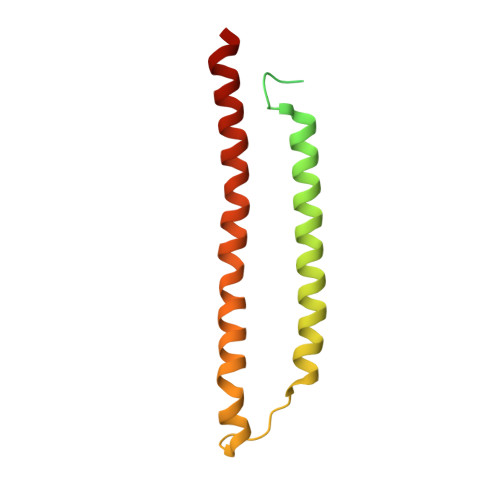
    Spindle- or lemon-shaped viruses infect archaea in diverse environments. Due to the highly pleomorphic nature of these virions, which can be found with cylindrical tails emanating from the spindle-shaped body, structural studies of these capsids have been challenging. We have determined the atomic structure of the capsid of Sulfolobus monocaudavirus 1, a virus that infects hosts living in nearly boiling acid. A highly hydrophobic protein, likely integrated into the host membrane before the virions assemble, forms 7 strands that slide past each other in both the tails and the spindle body. We observe the discrete steps that occur as the tail tubes expand, and these are due to highly conserved quasiequivalent interactions with neighboring subunits maintained despite significant diameter changes. Our results show how helical assemblies can vary their diameters, becoming nearly spherical to package a larger genome and suggest how all spindle-shaped viruses have evolved from archaeal rod-like viruses.


  • Organizational Affiliation
    • Department of Biochemistry and Molecular Genetics, University of Virginia, Charlottesville, VA 22903, USA.

Macromolecule Content 

  • Total Structure Weight: 16.18 kDa 
  • Atom Count: 686 
  • Modeled Residue Count: 94 
  • Deposited Residue Count: 157 
  • Unique protein chains: 1

Macromolecules

Find similar proteins by:|  3D Structure
Entity ID: 1
MoleculeChains  Sequence LengthOrganismDetailsImage
major capsid protein157Sulfolobus monocaudavirus SMV1Mutation(s): 0 
UniProt
Find proteins for A0ACD6B9J1 (Sulfolobus monocaudavirus SMV1)
Explore A0ACD6B9J1 
Go to UniProtKB:  A0ACD6B9J1
Entity Groups
Sequence Clusters30% Identity50% Identity70% Identity90% Identity95% Identity100% Identity
UniProt GroupA0ACD6B9J1
Sequence Annotations
Expand
Reference Sequence

Experimental Data & Validation

Experimental Data

  • Method: ELECTRON MICROSCOPY
  • Resolution: 4.00 Å
  • Aggregation State: FILAMENT 
  • Reconstruction Method: HELICAL 
EM Software:
TaskSoftware PackageVersion
MODEL REFINEMENTPHENIX

Structure Validation

View Full Validation Report


Entry History 

& Funding Information

Deposition Data


Funding OrganizationLocationGrant Number
National Institutes of Health/National Institute of General Medical Sciences (NIH/NIGMS)United StatesR35GM122510
National Institutes of Health/National Institute of General Medical Sciences (NIH/NIGMS)United StatesK99GM138756

Revision History  (Full details and data files)

  • Version 1.0: 2022-03-30
    Type: Initial release
  • Version 1.1: 2022-04-06
    Changes: Database references
  • Version 1.2: 2022-04-27
    Changes: Database references
  • Version 1.3: 2024-06-05
    Changes: Data collection
  • Version 1.4: 2025-05-14
    Changes: Data collection, Structure summary