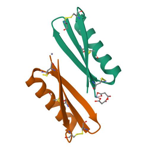
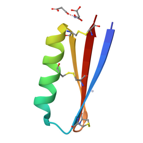
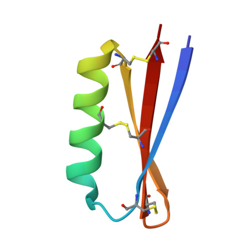
7RJF

MOPD-1 mutant-L47W


Experimental Data Snapshot

  • Method: X-RAY DIFFRACTION
  • Resolution: 1.80 Å
  • R-Value Free: 0.277 
  • R-Value Work: 0.232 
  • R-Value Observed: 0.237 

Starting Model: experimental
View more details

wwPDB Validation   3D Report Full Report


This is version 1.3 of the entry. See complete history


Literature

Rational Design of Potent Peptide Inhibitors of the PD-1:PD-L1 Interaction for Cancer Immunotherapy.

Yin, H.Zhou, X.Huang, Y.H.King, G.J.Collins, B.M.Gao, Y.Craik, D.J.Wang, C.K.

(2021) J Am Chem Soc 143: 18536-18547

  • DOI: https://doi.org/10.1021/jacs.1c08132
  • Primary Citation of Related Structures:  
    7RJF

  • PubMed Abstract: 

    Peptides have potential to be developed into immune checkpoint inhibitors, but the target interfaces are difficult to inhibit. Here, we explored an approach to mimic the binding surface of PD-1 to design inhibitors. Mimicking native PD-1 resulted in a mimetic with no activity. However, mimicking an affinity-optimized PD-1 resulted in the peptide mimetic MOPD-1 that displayed nanomolar affinity to PD-L1 and could inhibit PD-1:PD-L1 interactions in both protein- and cell-based assays. Mutagenesis and structural characterization using NMR spectroscopy and X-ray crystallography revealed that binding residues from the high affinity PD-1 are crucial for the bioactivity of MOPD-1. Furthermore, MOPD-1 was extremely stable in human serum and inhibited tumor growth in vivo , suggesting it has potential for use in cancer immunotherapy. The successful design of an inhibitor of PD-1:PD-L1 using the mimicry approach described herein illustrates the value of placing greater emphasis on optimizing the target interface before inhibitor design and is an approach that could have broader utility for the design of peptide inhibitors for other complex protein-protein interactions.


  • Organizational Affiliation

    Institute for Molecular Bioscience, The University of Queensland, Brisbane, Queensland 4072, Australia.


Macromolecules
Find similar proteins by:  (by identity cutoff)  |  3D Structure
Entity ID: 1
MoleculeChains Sequence LengthOrganismDetailsImage
[L47W]MOPD-1
A, B
47synthetic constructMutation(s): 0 
Entity Groups  
Sequence Clusters30% Identity50% Identity70% Identity90% Identity95% Identity100% Identity
Sequence Annotations
Expand
  • Reference Sequence
Experimental Data & Validation

Experimental Data

  • Method: X-RAY DIFFRACTION
  • Resolution: 1.80 Å
  • R-Value Free: 0.277 
  • R-Value Work: 0.232 
  • R-Value Observed: 0.237 
  • Space Group: P 41 21 2
Unit Cell:
Length ( Å )Angle ( ˚ )
a = 41.377α = 90
b = 41.377β = 90
c = 127.399γ = 90
Software Package:
Software NamePurpose
PHENIXrefinement
PDB_EXTRACTdata extraction
Aimlessdata scaling
PHASERphasing

Structure Validation

View Full Validation Report


Entry History & Funding Information

Deposition Data


Funding OrganizationLocationGrant Number
Australian Research Council (ARC)AustraliaCE200100012
National Health and Medical Research Council (NHMRC, Australia)AustraliaAPP1136021

Revision History  (Full details and data files)

  • Version 1.0: 2021-10-27
    Type: Initial release
  • Version 1.1: 2021-11-24
    Changes: Database references
  • Version 1.2: 2023-10-18
    Changes: Data collection, Refinement description
  • Version 1.3: 2024-10-23
    Changes: Structure summary