7RHR | pdb_00007rhr

Cryo-EM structure of Xenopus Patched-1 in nanodisc


Experimental Data Snapshot

  • Method: ELECTRON MICROSCOPY
  • Resolution: 3.00 Å
  • Aggregation State: PARTICLE 
  • Reconstruction Method: SINGLE PARTICLE 

wwPDB Validation 3D Report Full Report

Validation slider image for 7RHR

This is version 1.3 of the entry. See complete history

Literature

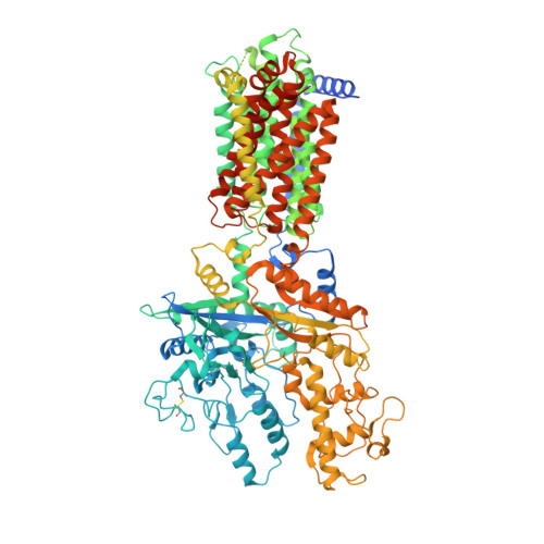
Structural basis for catalyzed assembly of the Sonic hedgehog-Patched1 signaling complex.

Huang, P.Wierbowski, B.M.Lian, T.Chan, C.Garcia-Linares, S.Jiang, J.Salic, A.

(2022) Dev Cell 57: 670-685.e8

  • DOI: https://doi.org/10.1016/j.devcel.2022.02.008
  • Primary Citation Related Structures: 
    7RHQ, 7RHR

  • PubMed Abstract: 

    The dually lipidated Sonic hedgehog (SHH) morphogen signals through the tumor suppressor membrane protein Patched1 (PTCH1) to activate the Hedgehog pathway, which is fundamental in development and cancer. SHH engagement with PTCH1 requires the GAS1 coreceptor, but the mechanism is unknown. We demonstrate a unique role for GAS1, catalyzing SHH-PTCH1 complex assembly in vertebrate cells by direct SHH transfer from the extracellular SCUBE2 carrier to PTCH1. Structure of the GAS1-SHH-PTCH1 transition state identifies how GAS1 recognizes the SHH palmitate and cholesterol modifications in modular fashion and how it facilitates lipid-dependent SHH handoff to PTCH1. Structure-guided experiments elucidate SHH movement from SCUBE2 to PTCH1, explain disease mutations, and demonstrate that SHH-induced PTCH1 dimerization causes its internalization from the cell surface. These results define how the signaling-competent SHH-PTCH1 complex assembles, the key step triggering the Hedgehog pathway, and provide a paradigm for understanding morphogen reception and its regulation.


  • Organizational Affiliation
    • Department of Cell Biology, Harvard Medical School, Boston, MA 02115, USA.

Macromolecule Content 

  • Total Structure Weight: 136.65 kDa 
  • Atom Count: 7,820 
  • Modeled Residue Count: 974 
  • Deposited Residue Count: 1,205 
  • Unique protein chains: 1

Macromolecules

Find similar proteins by:|  3D Structure
Entity ID: 1
MoleculeChains  Sequence LengthOrganismDetailsImage
Patched-11,205Xenopus calcaratusMutation(s): 0 
Membrane Entity: Yes 
UniProt
Find proteins for Q98SW6 (Xenopus laevis)
Explore Q98SW6 
Go to UniProtKB:  Q98SW6
Entity Groups
Sequence Clusters30% Identity50% Identity70% Identity90% Identity95% Identity100% Identity
UniProt GroupQ98SW6
Glycosylation
Glycosylation Sites: 3
Sequence Annotations
Expand
Reference Sequence

Oligosaccharides

Help  
Entity ID: 2
MoleculeChains Length2D Diagram GlycosylationD Interactions
2-acetamido-2-deoxy-beta-D-glucopyranose-(1-4)-2-acetamido-2-deoxy-beta-D-glucopyranose
B
2N-Glycosylation
Glycosylation Resources
GlyTouCan: G42666HT
GlyCosmos: G42666HT
GlyGen: G42666HT

Experimental Data & Validation

Experimental Data

  • Method: ELECTRON MICROSCOPY
  • Resolution: 3.00 Å
  • Aggregation State: PARTICLE 
  • Reconstruction Method: SINGLE PARTICLE 

Structure Validation

View Full Validation Report


Entry History 

& Funding Information

Deposition Data


Funding OrganizationLocationGrant Number
National Institutes of Health/National Institute of General Medical Sciences (NIH/NIGMS)United StatesR01GM122920

Revision History  (Full details and data files)

  • Version 1.0: 2022-03-02
    Type: Initial release
  • Version 1.1: 2022-03-09
    Changes: Database references
  • Version 1.2: 2022-03-23
    Changes: Database references
  • Version 1.3: 2024-10-09
    Changes: Data collection, Structure summary