7RF9 | pdb_00007rf9

O2-, PLP-dependent desaturase Plu4 intermediate-bound enzyme


Experimental Data Snapshot

  • Method: X-RAY DIFFRACTION
  • Resolution: 1.93 Å
  • R-Value Free: 
    0.194 (Depositor), 0.193 (DCC) 
  • R-Value Work: 
    0.151 (Depositor), 0.152 (DCC) 
  • R-Value Observed: 
    0.153 (Depositor) 

Starting Model: experimental
View more details

wwPDB Validation 3D Report Full Report

Validation slider image for 7RF9

Ligand Structure Quality Assessment 


This is version 1.1 of the entry. See complete history

Literature

A shared mechanistic pathway for pyridoxal phosphate-dependent arginine oxidases.

Hoffarth, E.R.Caddell Haatveit, K.Kuatsjah, E.MacNeil, G.A.Saroya, S.Walsby, C.J.Eltis, L.D.Houk, K.N.Garcia-Borras, M.Ryan, K.S.

(2021) Proc Natl Acad Sci U S A 118

  • DOI: https://doi.org/10.1073/pnas.2012591118
  • Primary Citation Related Structures: 
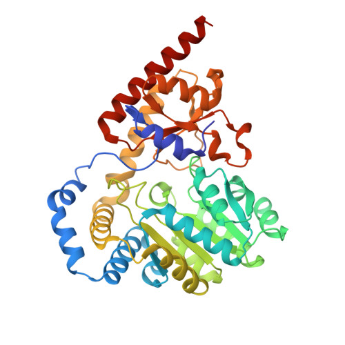
    7N79, 7RF9, 7RGB

  • PubMed Abstract: 

    The mechanism by which molecular oxygen is activated by the organic cofactor pyridoxal phosphate (PLP) for oxidation reactions remains poorly understood. Recent work has identified arginine oxidases that catalyze desaturation or hydroxylation reactions. Here, we investigate a desaturase from the Pseudoalteromonas luteoviolacea indolmycin pathway. Our work, combining X-ray crystallographic, biochemical, spectroscopic, and computational studies, supports a shared mechanism with arginine hydroxylases, involving two rounds of single-electron transfer to oxygen and superoxide rebound at the 4' carbon of the PLP cofactor. The precise positioning of a water molecule in the active site is proposed to control the final reaction outcome. This proposed mechanism provides a unified framework to understand how oxygen can be activated by PLP-dependent enzymes for oxidation of arginine and elucidates a shared mechanistic pathway and intertwined evolutionary history for arginine desaturases and hydroxylases.


  • Organizational Affiliation
    • Department of Chemistry, The University of British Columbia, Vancouver, BC V6T 1Z1, Canada.

Macromolecule Content 

  • Total Structure Weight: 182.92 kDa 
  • Atom Count: 13,549 
  • Modeled Residue Count: 1,513 
  • Deposited Residue Count: 1,556 
  • Unique protein chains: 1

Macromolecules

Find similar proteins by:|  3D Structure
Entity ID: 1
MoleculeChains  Sequence LengthOrganismDetailsImage
Aminotran_1_2 domain-containing protein
A, B, C, D
389Pseudoalteromonas luteoviolaceaMutation(s): 0 
Gene Names: JF50_03865
UniProt
Find proteins for A0A0C1MLE8 (Pseudoalteromonas luteoviolacea)
Explore A0A0C1MLE8 
Go to UniProtKB:  A0A0C1MLE8
Entity Groups
Sequence Clusters30% Identity50% Identity70% Identity90% Identity95% Identity100% Identity
UniProt GroupA0A0C1MLE8
Sequence Annotations
Expand
Reference Sequence

Small Molecules

Ligands 6 Unique
IDChains Name / Formula / InChI Key2D Diagram3D Interactions
EJ1
(Subject of Investigation/LOI)

Query on EJ1


Download:Ideal Coordinates CCD File
G [auth A],
GA [auth D],
S [auth B],
W [auth C]
(2E)-5-carbamimidamido-2-{[(Z)-{3-hydroxy-2-methyl-5-[(phosphonooxy)methyl]pyridin-4(1H)-ylidene}methyl]imino}pentanoic acid
C14 H22 N5 O7 P
RHCMSTSZICWJMS-GXVFZPQQSA-N
BTB

Query on BTB


Download:Ideal Coordinates CCD File
E [auth A],
EA [auth D],
H [auth A],
Q [auth B],
X [auth C]
2-[BIS-(2-HYDROXY-ETHYL)-AMINO]-2-HYDROXYMETHYL-PROPANE-1,3-DIOL
C8 H19 N O5
OWMVSZAMULFTJU-UHFFFAOYSA-N
AE3

Query on AE3


Download:Ideal Coordinates CCD File
Y [auth C]2-(2-ETHOXYETHOXY)ETHANOL
C6 H14 O3
XXJWXESWEXIICW-UHFFFAOYSA-N
GOL

Query on GOL


Download:Ideal Coordinates CCD File
I [auth A]GLYCEROL
C3 H8 O3
PEDCQBHIVMGVHV-UHFFFAOYSA-N
EDO

Query on EDO


Download:Ideal Coordinates CCD File
AA [auth C]
BA [auth C]
CA [auth C]
DA [auth C]
HA [auth D]
AA [auth C],
BA [auth C],
CA [auth C],
DA [auth C],
HA [auth D],
IA [auth D],
JA [auth D],
K [auth A],
KA [auth D],
L [auth A],
LA [auth D],
M [auth A],
MA [auth D],
N [auth A],
O [auth A],
P [auth A],
T [auth B],
U [auth B],
V [auth B]
1,2-ETHANEDIOL
C2 H6 O2
LYCAIKOWRPUZTN-UHFFFAOYSA-N
ACT

Query on ACT


Download:Ideal Coordinates CCD File
F [auth A],
FA [auth D],
J [auth A],
R [auth B],
Z [auth C]
ACETATE ION
C2 H3 O2
QTBSBXVTEAMEQO-UHFFFAOYSA-M

Experimental Data & Validation

Experimental Data

  • Method: X-RAY DIFFRACTION
  • Resolution: 1.93 Å
  • R-Value Free:  0.194 (Depositor), 0.193 (DCC) 
  • R-Value Work:  0.151 (Depositor), 0.152 (DCC) 
  • R-Value Observed: 0.153 (Depositor) 
Space Group: P 1 21 1
Unit Cell:
Length ( Å )Angle ( ˚ )
a = 74.922α = 90
b = 70.279β = 98.11
c = 138.568γ = 90
Software Package:
Software NamePurpose
Aimlessdata scaling
PHASERphasing
PHENIXrefinement
PDB_EXTRACTdata extraction
XDSdata reduction

Structure Validation

View Full Validation Report


Ligand Structure Quality Assessment 


Entry History 

& Funding Information

Deposition Data


Funding OrganizationLocationGrant Number
Natural Sciences and Engineering Research Council (NSERC, Canada)CanadaRGPIN-2016-03778

Revision History  (Full details and data files)

  • Version 1.0: 2021-10-06
    Type: Initial release
  • Version 1.1: 2023-10-18
    Changes: Data collection, Refinement description