7RDH | pdb_00007rdh

Crystal structure of the de novo designed binding protein H3mb in complex with the 1968 influenza A virus hemagglutinin


Experimental Data Snapshot

  • Method: X-RAY DIFFRACTION
  • Resolution: 2.75 Å
  • R-Value Free: 
    0.289 (Depositor), 0.302 (DCC) 
  • R-Value Work: 
    0.232 (Depositor), 0.249 (DCC) 
  • R-Value Observed: 
    0.235 (Depositor) 

Starting Model: experimental
View more details

wwPDB Validation 3D Report Full Report

Validation slider image for 7RDH

Ligand Structure Quality Assessment 


This is version 1.4 of the entry. See complete history

Literature

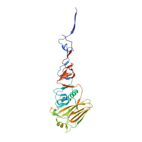
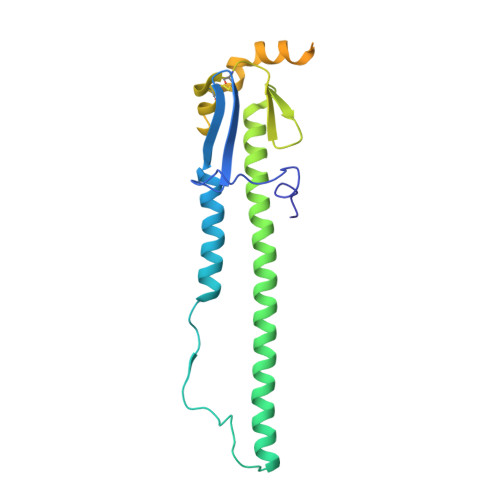
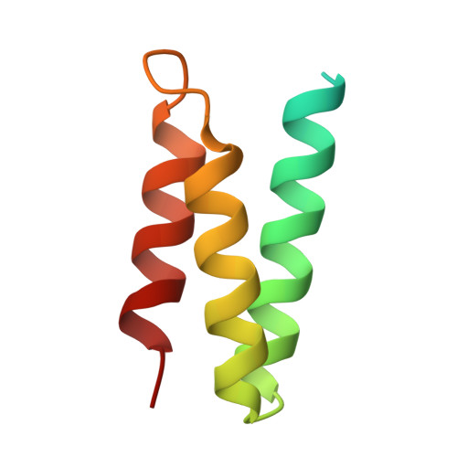
Design of protein-binding proteins from the target structure alone.

Cao, L.Coventry, B.Goreshnik, I.Huang, B.Sheffler, W.Park, J.S.Jude, K.M.Markovic, I.Kadam, R.U.Verschueren, K.H.G.Verstraete, K.Walsh, S.T.R.Bennett, N.Phal, A.Yang, A.Kozodoy, L.DeWitt, M.Picton, L.Miller, L.Strauch, E.M.DeBouver, N.D.Pires, A.Bera, A.K.Halabiya, S.Hammerson, B.Yang, W.Bernard, S.Stewart, L.Wilson, I.A.Ruohola-Baker, H.Schlessinger, J.Lee, S.Savvides, S.N.Garcia, K.C.Baker, D.

(2022) Nature 605: 551-560

  • DOI: https://doi.org/10.1038/s41586-022-04654-9
  • Primary Citation Related Structures: 
    7N1J, 7N1K, 7N3T, 7OPB, 7RDH, 7S5B

  • PubMed Abstract: 

    The design of proteins that bind to a specific site on the surface of a target protein using no information other than the three-dimensional structure of the target remains a challenge 1-5 . Here we describe a general solution to this problem that starts with a broad exploration of the vast space of possible binding modes to a selected region of a protein surface, and then intensifies the search in the vicinity of the most promising binding modes. We demonstrate the broad applicability of this approach through the de novo design of binding proteins to 12 diverse protein targets with different shapes and surface properties. Biophysical characterization shows that the binders, which are all smaller than 65 amino acids, are hyperstable and, following experimental optimization, bind their targets with nanomolar to picomolar affinities. We succeeded in solving crystal structures of five of the binder-target complexes, and all five closely match the corresponding computational design models. Experimental data on nearly half a million computational designs and hundreds of thousands of point mutants provide detailed feedback on the strengths and limitations of the method and of our current understanding of protein-protein interactions, and should guide improvements of both. Our approach enables the targeted design of binders to sites of interest on a wide variety of proteins for therapeutic and diagnostic applications.


  • Organizational Affiliation
    • Department of Biochemistry, University of Washington, Seattle, WA, USA.

Macromolecule Content 

  • Total Structure Weight: 211.39 kDa 
  • Atom Count: 12,887 
  • Modeled Residue Count: 1,591 
  • Deposited Residue Count: 1,840 
  • Unique protein chains: 3

Macromolecules

Find similar proteins by:|  3D Structure
Entity ID: 1
MoleculeChains  Sequence LengthOrganismDetailsImage
Hemagglutinin HA1 chain
A, C, E
323Influenza A virus (A/Hong Kong/1/1968(H3N2))Mutation(s): 0 
Gene Names: HA
UniProt
Find proteins for Q91MA7 (Influenza A virus (strain A/Hong Kong/1/1968 H3N2))
Explore Q91MA7 
Go to UniProtKB:  Q91MA7
Entity Groups
Sequence Clusters30% Identity50% Identity70% Identity90% Identity95% Identity100% Identity
UniProt GroupQ91MA7
Glycosylation
Glycosylation Sites: 4
Sequence Annotations
Expand
Reference Sequence
Find similar proteins by:|  3D Structure
Entity ID: 2
MoleculeChains  Sequence LengthOrganismDetailsImage
Hemagglutinin HA2 chain
B, D, F
239Influenza A virus (A/Hong Kong/1/1968(H3N2))Mutation(s): 1 
Gene Names: HA
UniProt
Find proteins for Q91MA7 (Influenza A virus (strain A/Hong Kong/1/1968 H3N2))
Explore Q91MA7 
Go to UniProtKB:  Q91MA7
Entity Groups
Sequence Clusters30% Identity50% Identity70% Identity90% Identity95% Identity100% Identity
UniProt GroupQ91MA7
Glycosylation
Glycosylation Sites: 1
Sequence Annotations
Expand
Reference Sequence
Find similar proteins by:|  3D Structure
Entity ID: 3
MoleculeChains  Sequence LengthOrganismDetailsImage
De novo designed protein H3mb
G, H
77Escherichia coliMutation(s): 0 

Oligosaccharides

Help  
Entity ID: 4
MoleculeChains Length2D Diagram GlycosylationD Interactions
2-acetamido-2-deoxy-beta-D-glucopyranose-(1-4)-2-acetamido-2-deoxy-beta-D-glucopyranose
I, J, K, M, O
2N-Glycosylation
Glycosylation Resources
GlyTouCan: G42666HT
GlyCosmos: G42666HT
GlyGen: G42666HT
Entity ID: 5
MoleculeChains Length2D Diagram GlycosylationD Interactions
beta-D-mannopyranose-(1-4)-2-acetamido-2-deoxy-beta-D-glucopyranose-(1-4)-2-acetamido-2-deoxy-beta-D-glucopyranose
L, N
3N-Glycosylation
Glycosylation Resources
GlyTouCan: G15407YE
GlyCosmos: G15407YE
GlyGen: G15407YE

Experimental Data & Validation

Experimental Data

  • Method: X-RAY DIFFRACTION
  • Resolution: 2.75 Å
  • R-Value Free:  0.289 (Depositor), 0.302 (DCC) 
  • R-Value Work:  0.232 (Depositor), 0.249 (DCC) 
  • R-Value Observed: 0.235 (Depositor) 
Space Group: P 1 21 1
Unit Cell:
Length ( Å )Angle ( ˚ )
a = 69.916α = 90
b = 240.845β = 117.304
c = 70.704γ = 90
Software Package:
Software NamePurpose
PHENIXrefinement
HKL-2000data reduction
HKL-2000data scaling
PHASERphasing

Structure Validation

View Full Validation Report


Ligand Structure Quality Assessment 


Entry History 

& Funding Information

Deposition Data


Funding OrganizationLocationGrant Number
National Institutes of Health/National Institute Of Allergy and Infectious Diseases (NIH/NIAID)United StatesR56 AI127371

Revision History  (Full details and data files)

  • Version 1.0: 2022-05-04
    Type: Initial release
  • Version 1.1: 2022-05-25
    Changes: Database references
  • Version 1.2: 2022-06-01
    Changes: Database references
  • Version 1.3: 2023-10-18
    Changes: Data collection, Refinement description
  • Version 1.4: 2024-11-06
    Changes: Structure summary