7RCP

GltPh mutant (S279E/D405N) in complex with aspartate and sodium ions


Experimental Data Snapshot

  • Method: ELECTRON MICROSCOPY
  • Resolution: 2.20 Å
  • Aggregation State: PARTICLE 
  • Reconstruction Method: SINGLE PARTICLE 

wwPDB Validation   3D Report Full Report


This is version 1.2 of the entry. See complete history


Literature

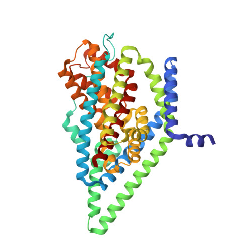
The archaeal glutamate transporter homologue GltPh shows heterogeneous substrate binding.

Reddy, K.D.Ciftci, D.Scopelliti, A.J.Boudker, O.

(2022) J Gen Physiol 154

  • DOI: https://doi.org/10.1085/jgp.202213131
  • Primary Citation of Related Structures:  
    7RCP

  • PubMed Abstract: 

    Integral membrane glutamate transporters couple the concentrative substrate transport to ion gradients. There is a wealth of structural and mechanistic information about this protein family. Recent studies of an archaeal homologue, GltPh, revealed transport rate heterogeneity, which is inconsistent with simple kinetic models; however, its structural and mechanistic determinants remain undefined. Here, we demonstrate that in a mutant GltPh, which exclusively populates the outward-facing state, at least two substates coexist in slow equilibrium, binding the substrate with different apparent affinities. Wild type GltPh shows similar binding properties, and modulation of the substate equilibrium correlates with transport rates. The low-affinity substate of the mutant is transient following substrate binding. Consistently, cryo-EM on samples frozen within seconds after substrate addition reveals the presence of structural classes with perturbed helical packing of the extracellular half of the transport domain in regions adjacent to the binding site. By contrast, an equilibrated structure does not show such classes. The structure at 2.2-Å resolution details a pattern of waters in the intracellular half of the domain and resolves classes with subtle differences in the substrate-binding site. We hypothesize that the rigid cytoplasmic half of the domain mediates substrate and ion recognition and coupling, whereas the extracellular labile half sets the affinity and dynamic properties.


  • Organizational Affiliation

    Department of Physiology and Biophysics, Weill Cornell Medicine, New York, NY.


Macromolecules
Find similar proteins by:  (by identity cutoff)  |  3D Structure
Entity ID: 1
MoleculeChains Sequence LengthOrganismDetailsImage
Glutamate transporter homolog
A, B, C
415Pyrococcus horikoshii OT3Mutation(s): 2 
Gene Names: PH1295
Membrane Entity: Yes 
UniProt
Find proteins for O59010 (Pyrococcus horikoshii (strain ATCC 700860 / DSM 12428 / JCM 9974 / NBRC 100139 / OT-3))
Explore O59010 
Go to UniProtKB:  O59010
Entity Groups  
Sequence Clusters30% Identity50% Identity70% Identity90% Identity95% Identity100% Identity
UniProt GroupO59010
Sequence Annotations
Expand
  • Reference Sequence
Small Molecules
Ligands 2 Unique
IDChains Name / Formula / InChI Key2D Diagram3D Interactions
ASP (Subject of Investigation/LOI)
Query on ASP

Download Ideal Coordinates CCD File 
D [auth A],
G [auth B],
J [auth C]
ASPARTIC ACID
C4 H7 N O4
CKLJMWTZIZZHCS-REOHCLBHSA-N
NA (Subject of Investigation/LOI)
Query on NA

Download Ideal Coordinates CCD File 
E [auth A]
F [auth A]
H [auth B]
I [auth B]
K [auth C]
E [auth A],
F [auth A],
H [auth B],
I [auth B],
K [auth C],
L [auth C]
SODIUM ION
Na
FKNQFGJONOIPTF-UHFFFAOYSA-N
Experimental Data & Validation

Experimental Data

  • Method: ELECTRON MICROSCOPY
  • Resolution: 2.20 Å
  • Aggregation State: PARTICLE 
  • Reconstruction Method: SINGLE PARTICLE 

Structure Validation

View Full Validation Report


Entry History & Funding Information

Deposition Data


Funding OrganizationLocationGrant Number
National Institutes of Health/National Institute of Neurological Disorders and Stroke (NIH/NINDS)United StatesR01NS064357
National Institutes of Health/National Institute of Neurological Disorders and Stroke (NIH/NINDS)United StatesR37NS085318
National Institutes of Health/National Institute of Neurological Disorders and Stroke (NIH/NINDS)United StatesNS102325

Revision History  (Full details and data files)

  • Version 1.0: 2022-04-20
    Type: Initial release
  • Version 1.1: 2022-11-02
    Changes: Database references
  • Version 1.2: 2024-06-05
    Changes: Data collection