7RBW | pdb_00007rbw

Structure of Biliverdin-binding Serpin of Boana punctata (polka-dot tree frog)


Experimental Data Snapshot

  • Method: X-RAY DIFFRACTION
  • Resolution: 2.05 Å
  • R-Value Free: 
    0.237 (Depositor), 0.266 (DCC) 
  • R-Value Work: 
    0.199 (Depositor) 
  • R-Value Observed: 
    0.201 (Depositor) 

Starting Model: experimental
View more details

wwPDB Validation 3D Report Full Report

Validation slider image for 7RBW

Ligand Structure Quality Assessment 


This is version 1.3 of the entry. See complete history

Literature

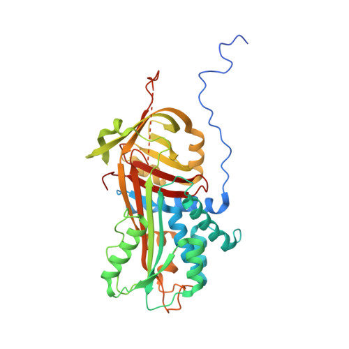
Structural and Functional Characterization of a Biliverdin-Binding Near-Infrared Fluorescent Protein From the Serpin Superfamily.

Manoilov, K.Y.Ghosh, A.Almo, S.C.Verkhusha, V.V.

(2021) J Mol Biology 434: 167359-167359

  • DOI: https://doi.org/10.1016/j.jmb.2021.167359
  • Primary Citation Related Structures: 
    7RBW

  • PubMed Abstract: 

    Biliverdin-binding serpins (BBSs) are proteins that are responsible for coloration in amphibians and fluoresce in the near-infrared (NIR) spectral region. Here we produced the first functional recombinant BBS of the polka-dot treefrog Boana punctata (BpBBS), assembled with its biliverdin (BV) chromophore, and report its biochemical and photochemical characterization. We determined the crystal structure of BpBBS at 2.05 Å resolution, which demonstrated its structural homology to the mammalian protease inhibitor alpha-1-antitrypsin. BV interaction with BpBBS was studied and it was found that the N-terminal polypeptide (residues 19-50) plays a critical role in the BV binding. By comparing BpBBS with the available NIR fluorescent proteins and expressing it in mammalian cells, we demonstrated its potential as a NIR imaging probe. These results provide insight into the non-inhibitory function of serpins, provide a basis for improving their performance in mammalian cells, and suggest possible paths for the development of BBS-based fluorescent probes.


  • Organizational Affiliation
    • Department of Anatomy and Structural Biology, Albert Einstein College of Medicine, Bronx, NY 10461, USA.

Macromolecule Content 

  • Total Structure Weight: 95.82 kDa 
  • Atom Count: 6,390 
  • Modeled Residue Count: 760 
  • Deposited Residue Count: 822 
  • Unique protein chains: 1

Macromolecules

Find similar proteins by:|  3D Structure
Entity ID: 1
MoleculeChains  Sequence LengthOrganismDetailsImage
Biliverdin bindin serpin
A, B
411Boana punctataMutation(s): 0 
Gene Names: BBS
UniProt
Find proteins for A0A7D7FB99 (Boana punctata)
Explore A0A7D7FB99 
Go to UniProtKB:  A0A7D7FB99
Entity Groups
Sequence Clusters30% Identity50% Identity70% Identity90% Identity95% Identity100% Identity
UniProt GroupA0A7D7FB99
Sequence Annotations
Expand
Reference Sequence

Experimental Data & Validation

Experimental Data

  • Method: X-RAY DIFFRACTION
  • Resolution: 2.05 Å
  • R-Value Free:  0.237 (Depositor), 0.266 (DCC) 
  • R-Value Work:  0.199 (Depositor) 
  • R-Value Observed: 0.201 (Depositor) 
Space Group: P 21 21 21
Unit Cell:
Length ( Å )Angle ( ˚ )
a = 72.069α = 90
b = 75.91β = 90
c = 143.751γ = 90
Software Package:
Software NamePurpose
PHENIXrefinement
MOSFLMdata reduction
Aimlessdata scaling
PHASERphasing
PDB_EXTRACTdata extraction

Structure Validation

View Full Validation Report


Ligand Structure Quality Assessment 


Entry History 

& Funding Information

Deposition Data


Funding OrganizationLocationGrant Number
National Institutes of Health/National Institute of General Medical Sciences (NIH/NIGMS)United States5R35GM122567

Revision History  (Full details and data files)

  • Version 1.0: 2021-11-24
    Type: Initial release
  • Version 1.1: 2021-12-01
    Changes: Database references
  • Version 1.2: 2021-12-08
    Changes: Database references
  • Version 1.3: 2023-10-18
    Changes: Data collection, Refinement description