7R8L | pdb_00007r8l

Structure of the SARS-CoV-2 RBD in complex with neutralizing antibody C099 and CR3022


Experimental Data Snapshot

  • Method: X-RAY DIFFRACTION
  • Resolution: 2.60 Å
  • R-Value Free: 
    0.229 (Depositor), 0.229 (DCC) 
  • R-Value Work: 
    0.180 (Depositor), 0.179 (DCC) 
  • R-Value Observed: 
    0.182 (Depositor) 

Starting Model: experimental
View more details

wwPDB Validation 3D Report Full Report

Validation slider image for 7R8L

This is version 1.4 of the entry. See complete history

Literature

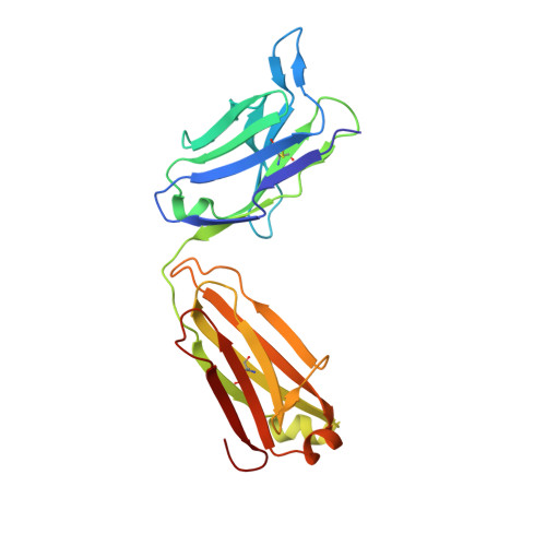
Affinity maturation of SARS-CoV-2 neutralizing antibodies confers potency, breadth, and resilience to viral escape mutations.

Muecksch, F.Weisblum, Y.Barnes, C.O.Schmidt, F.Schaefer-Babajew, D.Wang, Z.C Lorenzi, J.C.Flyak, A.I.DeLaitsch, A.T.Huey-Tubman, K.E.Hou, S.Schiffer, C.A.Gaebler, C.Da Silva, J.Poston, D.Finkin, S.Cho, A.Cipolla, M.Oliveira, T.Y.Millard, K.G.Ramos, V.Gazumyan, A.Rutkowska, M.Caskey, M.Nussenzweig, M.C.Bjorkman, P.J.Hatziioannou, T.Bieniasz, P.D.

(2021) Immunity 54: 1853-1868.e7

  • DOI: https://doi.org/10.1016/j.immuni.2021.07.008
  • Primary Citation Related Structures: 
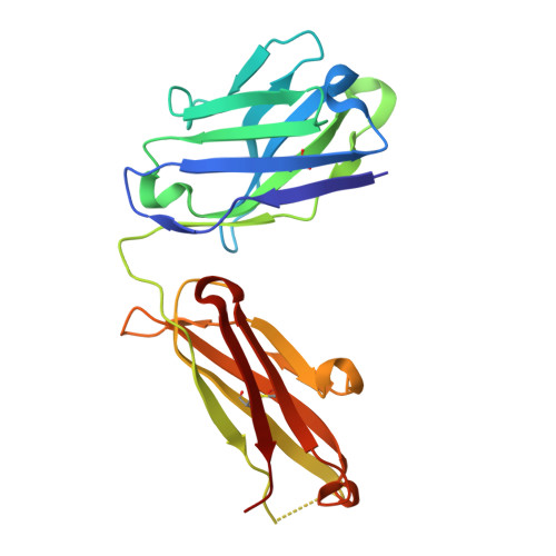
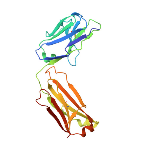
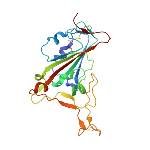
    7N3E, 7N3F, 7N3G, 7N3H, 7N3I, 7R8L, 7R8M, 7R8N, 7R8O

  • PubMed Abstract: 

    Antibodies elicited by infection accumulate somatic mutations in germinal centers that can increase affinity for cognate antigens. We analyzed 6 independent groups of clonally related severe acute respiratory syndrome-coronavirus-2 (SARS-CoV-2) Spike receptor-binding domain (RBD)-specific antibodies from 5 individuals shortly after infection and later in convalescence to determine the impact of maturation over months. In addition to increased affinity and neutralization potency, antibody evolution changed the mutational pathways for the acquisition of viral resistance and restricted neutralization escape options. For some antibodies, maturation imposed a requirement for multiple substitutions to enable escape. For certain antibodies, affinity maturation enabled the neutralization of circulating SARS-CoV-2 variants of concern and heterologous sarbecoviruses. Antibody-antigen structures revealed that these properties resulted from substitutions that allowed additional variability at the interface with the RBD. These findings suggest that increasing antibody diversity through prolonged or repeated antigen exposure may improve protection against diversifying SARS-CoV-2 populations, and perhaps against other pandemic threat coronaviruses.


  • Organizational Affiliation
    • Laboratory of Retrovirology, The Rockefeller University, New York, NY 10065, USA.

Macromolecule Content 

  • Total Structure Weight: 116.44 kDa 
  • Atom Count: 8,251 
  • Modeled Residue Count: 1,064 
  • Deposited Residue Count: 1,069 
  • Unique protein chains: 5

Macromolecules

Find similar proteins by:|  3D Structure
Entity ID: 1
MoleculeChains  Sequence LengthOrganismDetailsImage
C099 Fab Heavy ChainA [auth H]218Homo sapiensMutation(s): 0 
Find similar proteins by:|  3D Structure
Entity ID: 2
MoleculeChains  Sequence LengthOrganismDetailsImage
C099 Fab Light ChainB [auth L]213Homo sapiensMutation(s): 0 
Find similar proteins by:|  3D Structure
Entity ID: 3
MoleculeChains  Sequence LengthOrganismDetailsImage
Spike protein S1C [auth E]195Severe acute respiratory syndrome coronavirus 2Mutation(s): 0 
Gene Names: S2
UniProt
Find proteins for P0DTC2 (Severe acute respiratory syndrome coronavirus 2)
Explore P0DTC2 
Go to UniProtKB:  P0DTC2
Entity Groups
Sequence Clusters30% Identity50% Identity70% Identity90% Identity95% Identity100% Identity
UniProt GroupP0DTC2
Glycosylation
Glycosylation Sites: 1Go to GlyGen: P0DTC2-1
Sequence Annotations
Expand
Reference Sequence
Find similar proteins by:|  3D Structure
Entity ID: 4
MoleculeChains  Sequence LengthOrganismDetailsImage
CR3022 Fab heavy chainD [auth C]222Homo sapiensMutation(s): 0 
Find similar proteins by:|  3D Structure
Entity ID: 5
MoleculeChains  Sequence LengthOrganismDetailsImage
CR3022 Fab light chainE [auth D]221Homo sapiensMutation(s): 0 

Oligosaccharides

Help  
Entity ID: 6
MoleculeChains Length2D Diagram GlycosylationD Interactions
beta-D-mannopyranose-(1-4)-2-acetamido-2-deoxy-beta-D-glucopyranose-(1-4)-[alpha-L-fucopyranose-(1-6)]2-acetamido-2-deoxy-beta-D-glucopyranoseF [auth A]4N-Glycosylation
Glycosylation Resources
GlyTouCan: G32152BH
GlyCosmos: G32152BH
GlyGen: G32152BH

Experimental Data & Validation

Experimental Data

  • Method: X-RAY DIFFRACTION
  • Resolution: 2.60 Å
  • R-Value Free:  0.229 (Depositor), 0.229 (DCC) 
  • R-Value Work:  0.180 (Depositor), 0.179 (DCC) 
  • R-Value Observed: 0.182 (Depositor) 
Space Group: P 41 21 2
Unit Cell:
Length ( Å )Angle ( ˚ )
a = 109.947α = 90
b = 109.947β = 90
c = 228.554γ = 90
Software Package:
Software NamePurpose
PHENIXrefinement
Aimlessdata scaling
XDSdata reduction
PHASERphasing

Structure Validation

View Full Validation Report


Entry History 

& Funding Information

Deposition Data


Funding OrganizationLocationGrant Number
National Institutes of Health/National Institute Of Allergy and Infectious Diseases (NIH/NIAID)United StatesP01-AI138398

Revision History  (Full details and data files)

  • Version 1.0: 2021-08-04
    Type: Initial release
  • Version 1.1: 2021-08-11
    Changes: Database references
  • Version 1.2: 2021-08-25
    Changes: Database references
  • Version 1.3: 2023-10-18
    Changes: Data collection, Database references, Refinement description
  • Version 1.4: 2024-11-06
    Changes: Structure summary