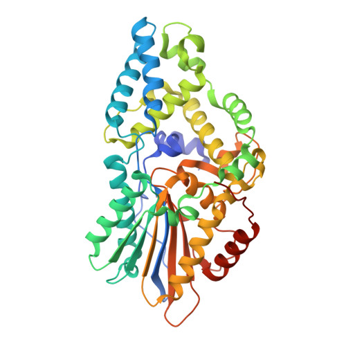
Crystal Structure of Prevotella sp. CAG:617 Multiple Inositol Polyphosphate Phosphatase, complex with myo-inositol hexakissulfate
Acquistapace, I.M., Brearley, C.A., Hemmings, A.M.To be published.
Experimental Data Snapshot
Starting Model: experimental
View more details
| Ligands 2 Unique | |||||
|---|---|---|---|---|---|
| ID | Chains | Name / Formula / InChI Key | 2D Diagram | 3D Interactions | |
| IHS (Subject of Investigation/LOI) Download:Ideal Coordinates CCD File | G [auth A] I [auth B] J [auth C] K [auth D] L [auth E] | D-MYO-INOSITOL-HEXASULPHATE C6 H12 O24 S6 NBTMNFYXJYCQHQ-GPIVLXJGSA-N |  | ||
| SO4 Download:Ideal Coordinates CCD File | H [auth A] | SULFATE ION O4 S QAOWNCQODCNURD-UHFFFAOYSA-L |  | ||
| Length ( Å ) | Angle ( ˚ ) |
|---|---|
| a = 145.14 | α = 90 |
| b = 263.63 | β = 90 |
| c = 85.03 | γ = 90 |
| Software Name | Purpose |
|---|---|
| GDA | data collection |
| PHENIX | refinement |
| xia2 | data reduction |
| Aimless | data scaling |
| PHASER | phasing |
| Funding Organization | Location | Grant Number |
|---|---|---|
| Biotechnology and Biological Sciences Research Council (BBSRC) | United Kingdom | BB/M022978/1 |