7R1Q | pdb_00007r1q

X-ray structure of the adduct formed upon reaction of the gold(I) N-heterocyclic carbene complex Au2 with lysozyme


Experimental Data Snapshot

  • Method: X-RAY DIFFRACTION
  • Resolution: 1.10 Å
  • R-Value Free: 
    0.170 (Depositor), 0.170 (DCC) 
  • R-Value Work: 
    0.132 (Depositor), 0.132 (DCC) 

Starting Model: experimental
View more details

wwPDB Validation 3D Report Full Report

Validation slider image for 7R1Q

This is version 1.3 of the entry. See complete history

Literature

Halo complexes of gold(I) containing glycoconjugate carbene ligands: synthesis, characterization, cytotoxicity and interaction with proteins and DNA model systems.

Annunziata, A.Ferraro, G.Cucciolito, M.E.Imbimbo, P.Tuzi, A.Monti, D.M.Merlino, A.Ruffo, F.

(2022) Dalton Trans 51: 10475-10485

  • DOI: https://doi.org/10.1039/d2dt00423b
  • Primary Citation Related Structures: 
    7R1P, 7R1Q

  • PubMed Abstract: 

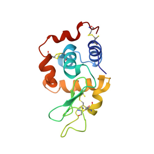
    New neutral carbene complexes of gold(I) [Au(Im-Me)X] (X = Cl, Au1; X = Br, Au2; X = I, Au3) have been synthesized and fully characterized by different techniques, including NMR and UV-vis absorption spectroscopy and single crystal X-ray diffraction. The carbene ligand Im-Me is decorated with a glucoside fragment via a triazole linker, obtainable through a click chemistry reaction. The compounds retain the Au-NHC fragment in aqueous solvents, and an equilibrium between the neutral halo- and the cationic di-carbene form [Au(Im-Me) 2 ] + is observed, whose extent follows the trend Au1 < Au2 < Au3. Cytotoxicity studies on two cancer and two non-tumorigenic cell lines reflect the solution behavior, as a certain difference among the complexes was disclosed, with the iodo complex Au3 being more active and selective. The compounds interact with both DNA and protein model systems. The X-ray structure of the adduct formed upon the reaction of Au1 with bovine pancreatic ribonuclease (RNase A) reveals Au binding at the side chain of His105 of both protein molecules A and B of the asymmetric unit. The binding of gold atoms at both the nitrogen atoms of the imidazole ring of His15 and at the N-terminal tail has been found in the adduct formed with hen egg white lysozyme.


  • Organizational Affiliation
    • Dipartimento di Scienze Chimiche, Università di Napoli Federico II, Complesso Universitario di Monte Sant'Angelo, Via Cintia 21, 80126, Napoli, Italy. dariamaria.monti@unina.it.

Macromolecule Content 

  • Total Structure Weight: 15 kDa 
  • Atom Count: 1,370 
  • Modeled Residue Count: 129 
  • Deposited Residue Count: 129 
  • Unique protein chains: 1

Macromolecules

Find similar proteins by:|  3D Structure
Entity ID: 1
MoleculeChains  Sequence LengthOrganismDetailsImage
LysozymeA [auth AAA]129Gallus gallusMutation(s): 0 
EC: 3.2.1.17
UniProt
Find proteins for P00698 (Gallus gallus)
Explore P00698 
Go to UniProtKB:  P00698
Entity Groups
Sequence Clusters30% Identity50% Identity70% Identity90% Identity95% Identity100% Identity
UniProt GroupP00698
Sequence Annotations
Expand
Reference Sequence

Experimental Data & Validation

Experimental Data

  • Method: X-RAY DIFFRACTION
  • Resolution: 1.10 Å
  • R-Value Free:  0.170 (Depositor), 0.170 (DCC) 
  • R-Value Work:  0.132 (Depositor), 0.132 (DCC) 
Space Group: P 43 21 2
Unit Cell:
Length ( Å )Angle ( ˚ )
a = 77α = 90
b = 77β = 90
c = 37.27γ = 90
Software Package:
Software NamePurpose
REFMACrefinement
autoPROCdata reduction
autoPROCdata scaling
PHASERphasing

Structure Validation

View Full Validation Report


Entry History 

& Funding Information

Deposition Data


Funding OrganizationLocationGrant Number
Not funded--

Revision History  (Full details and data files)

  • Version 1.0: 2022-07-13
    Type: Initial release
  • Version 1.1: 2022-07-20
    Changes: Database references
  • Version 1.2: 2024-01-31
    Changes: Data collection, Refinement description
  • Version 1.3: 2024-11-13
    Changes: Structure summary