7R0L | pdb_00007r0l

Structure of the FK1 domain of the FKBP51 G64S variant in complex with SAFit1


Experimental Data Snapshot

  • Method: X-RAY DIFFRACTION
  • Resolution: 1.10 Å
  • R-Value Free: 
    0.171 (Depositor), 0.170 (DCC) 
  • R-Value Work: 
    0.143 (Depositor), 0.141 (DCC) 

Starting Model: experimental
View more details

wwPDB Validation 3D Report Full Report

Validation slider image for 7R0L

Ligand Structure Quality Assessment 


This is version 1.2 of the entry. See complete history

Literature

Binding pocket stabilization by high-throughput screening of yeast display libraries.

Lerma Romero, J.A.Meyners, C.Christmann, A.Reinbold, L.M.Charalampidou, A.Hausch, F.Kolmar, H.

(2022) Front Mol Biosci 9: 1023131-1023131

  • DOI: https://doi.org/10.3389/fmolb.2022.1023131
  • Primary Citation Related Structures: 
    7R0L, 8BA6, 8BAJ

  • PubMed Abstract: 

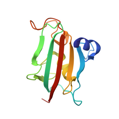
    Protein dynamics have a great influence on the binding pockets of some therapeutic targets. Flexible protein binding sites can result in transient binding pocket formation which might have a negative impact on drug screening efforts. Here, we describe a protein engineering strategy with FK506-binding protein 51 (FKBP51) as a model protein, which is a promising target for stress-related disorders. High-throughput screening of yeast display libraries of FKBP51 resulted in the identification of variants exhibiting higher affinity binding of conformation-specific FKBP51 selective inhibitors. The gene libraries of a random mutagenesis and site saturation mutagenesis of the FK1 domain of FKBP51 encoding sequence were used to create a yeast surface display library. Fluorescence-activated cell sorting for FKBP51 variants that bind conformation-specific fluorescently labeled ligands with high affinity allowed for the identification of 15 different protein variants with improved binding to either, or both FKBP51-specific ligands used in the screening, with improved affinities up to 34-fold compared to the wild type. These variants will pave the way to a better understanding of the conformational flexibility of the FKBP51 binding pocket and may enable the isolation of new selective ligands that preferably and selectively bind the active site of the protein in its open conformation state.


  • Organizational Affiliation
    • Institute for Organic Chemistry and Biochemistry, Technical University of Darmstadt, Darmstadt, Germany.

Macromolecule Content 

  • Total Structure Weight: 15.2 kDa 
  • Atom Count: 1,222 
  • Modeled Residue Count: 128 
  • Deposited Residue Count: 128 
  • Unique protein chains: 1

Macromolecules

Find similar proteins by:|  3D Structure
Entity ID: 1
MoleculeChains  Sequence LengthOrganismDetailsImage
Peptidyl-prolyl cis-trans isomerase FKBP5128Homo sapiensMutation(s): 4 
Gene Names: FKBP5AIG6FKBP51
EC: 5.2.1.8
UniProt & NIH Common Fund Data Resources
Find proteins for Q13451 (Homo sapiens)
Explore Q13451 
Go to UniProtKB:  Q13451
PHAROS:  Q13451
GTEx:  ENSG00000096060 
Entity Groups
Sequence Clusters30% Identity50% Identity70% Identity90% Identity95% Identity100% Identity
UniProt GroupQ13451
Sequence Annotations
Expand
Reference Sequence

Small Molecules

Ligands 3 Unique
IDChains Name / Formula / InChI Key2D Diagram3D Interactions
GY1
(Subject of Investigation/LOI)

Query on GY1


Download:Ideal Coordinates CCD File
B [auth A]2-[3-[(1~{R})-1-[(2~{S})-1-[(2~{S})-2-cyclohexyl-2-(3,4,5-trimethoxyphenyl)ethanoyl]piperidin-2-yl]carbonyloxy-3-(3,4-dimethoxyphenyl)propyl]phenoxy]ethanoic acid
C42 H53 N O11
OEQZPFWOEOOISR-AKTKKGGCSA-N
EPE

Query on EPE


Download:Ideal Coordinates CCD File
C [auth A]4-(2-HYDROXYETHYL)-1-PIPERAZINE ETHANESULFONIC ACID
C8 H18 N2 O4 S
JKMHFZQWWAIEOD-UHFFFAOYSA-N
GOL

Query on GOL


Download:Ideal Coordinates CCD File
D [auth A],
E [auth A]
GLYCEROL
C3 H8 O3
PEDCQBHIVMGVHV-UHFFFAOYSA-N
Binding Affinity Annotations 
IDSourceBinding Affinity
GY1 BindingDB:  7R0L Ki: min: 4, max: 6 (nM) from 2 assay(s)
Kd: 9.8 (nM) from 1 assay(s)

Experimental Data & Validation

Experimental Data

  • Method: X-RAY DIFFRACTION
  • Resolution: 1.10 Å
  • R-Value Free:  0.171 (Depositor), 0.170 (DCC) 
  • R-Value Work:  0.143 (Depositor), 0.141 (DCC) 
Space Group: P 21 21 21
Unit Cell:
Length ( Å )Angle ( ˚ )
a = 45.106α = 90
b = 48.592β = 90
c = 57.191γ = 90
Software Package:
Software NamePurpose
REFMACrefinement
REFMACrefinement
XDSdata reduction
Aimlessdata scaling
PHASERphasing

Structure Validation

View Full Validation Report


Ligand Structure Quality Assessment 


Entry History 

& Funding Information

Deposition Data


Funding OrganizationLocationGrant Number
Hessian Ministry of Science, Higher Education and Art (HMWK)Germany--

Revision History  (Full details and data files)

  • Version 1.0: 2022-11-16
    Type: Initial release
  • Version 1.1: 2022-12-07
    Changes: Database references
  • Version 1.2: 2024-01-31
    Changes: Data collection, Refinement description