7R03 | pdb_00007r03

Neurofibromin occluded conformation


Experimental Data Snapshot

  • Method: ELECTRON MICROSCOPY
  • Resolution: 3.60 Å
  • Aggregation State: PARTICLE 
  • Reconstruction Method: SINGLE PARTICLE 

Starting Models: experimental
View more details

wwPDB Validation 3D Report Full Report

Validation slider image for 7R03

This is version 1.3 of the entry. See complete history

Literature

Structural basis of activation of the tumor suppressor protein neurofibromin.

Chaker-Margot, M.Werten, S.Dunzendorfer-Matt, T.Lechner, S.Ruepp, A.Scheffzek, K.Maier, T.

(2022) Mol Cell 82: 1288

  • DOI: https://doi.org/10.1016/j.molcel.2022.03.011
  • Primary Citation Related Structures: 
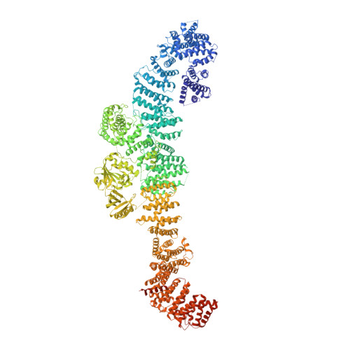
    7R03, 7R04

  • PubMed Abstract: 

    Mutations in the NF1 gene cause the familial genetic disease neurofibromatosis type I, as well as predisposition to cancer. The NF1 gene product, neurofibromin, is a GTPase-activating protein and acts as a tumor suppressor by negatively regulating the small GTPase, Ras. However, structural insights into neurofibromin activation remain incompletely defined. Here, we provide cryoelectron microscopy (cryo-EM) structures that reveal an extended neurofibromin homodimer in two functional states: an auto-inhibited state with occluded Ras-binding site and an asymmetric open state with an exposed Ras-binding site. Mechanistically, the transition to the active conformation is stimulated by nucleotide binding, which releases a lock that tethers the catalytic domain to an extended helical repeat scaffold in the occluded state. Structure-guided mutational analysis supports functional relevance of allosteric control. Disease-causing mutations are mapped and primarily impact neurofibromin stability. Our findings suggest a role for nucleotides in neurofibromin regulation and may lead to therapeutic modulation of Ras signaling.


  • Organizational Affiliation
    • Biozentrum, University of Basel, 4056 Basel, Switzerland.

Macromolecule Content 

  • Total Structure Weight: 634.82 kDa 
  • Atom Count: 35,621 
  • Modeled Residue Count: 4,487 
  • Deposited Residue Count: 5,636 
  • Unique protein chains: 1

Macromolecules

Find similar proteins by:|  3D Structure
Entity ID: 1
MoleculeChains  Sequence LengthOrganismDetailsImage
Isoform I of Neurofibromin
A, B
2,818Homo sapiensMutation(s): 0 
Gene Names: NF1
UniProt & NIH Common Fund Data Resources
Find proteins for P21359 (Homo sapiens)
Explore P21359 
Go to UniProtKB:  P21359
PHAROS:  P21359
GTEx:  ENSG00000196712 
Entity Groups
Sequence Clusters30% Identity50% Identity70% Identity90% Identity95% Identity100% Identity
UniProt GroupP21359
Sequence Annotations
Expand
Reference Sequence

Experimental Data & Validation

Experimental Data

  • Method: ELECTRON MICROSCOPY
  • Resolution: 3.60 Å
  • Aggregation State: PARTICLE 
  • Reconstruction Method: SINGLE PARTICLE 
EM Software:
TaskSoftware PackageVersion
RECONSTRUCTIONcryoSPARC2.14
MODEL REFINEMENTPHENIX1.19

Structure Validation

View Full Validation Report


Entry History 

& Funding Information

Deposition Data


Funding OrganizationLocationGrant Number
Human Frontier Science Program (HFSP)Switzerland--

Revision History  (Full details and data files)

  • Version 1.0: 2022-03-30
    Type: Initial release
  • Version 1.1: 2022-04-13
    Changes: Database references
  • Version 1.2: 2022-04-20
    Changes: Database references
  • Version 1.3: 2024-07-17
    Changes: Data collection, Refinement description