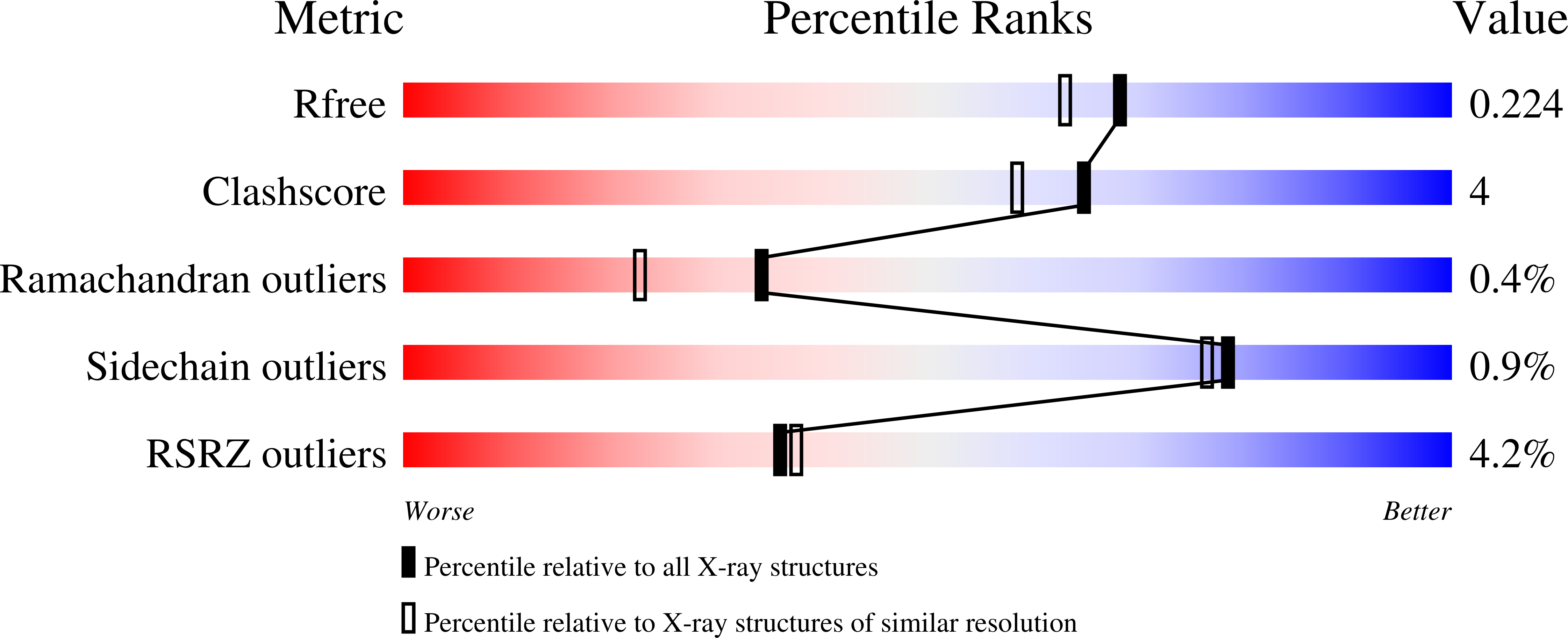
7QZ2

Crystal structure of GacS D1 domain in complex with BeF3-


Experimental Data Snapshot

  • Method: X-RAY DIFFRACTION
  • Resolution: 1.87 Å
  • R-Value Free: 0.213 
  • R-Value Work: 0.168 
  • R-Value Observed: 0.171 

Starting Model: experimental
View more details

wwPDB Validation   3D Report Full Report


This is version 1.2 of the entry. See complete history


Literature

Insights into the atypical autokinase activity of the Pseudomonas aeruginosa GacS histidine kinase and its interaction with RetS.

Fadel, F.Bassim, V.Francis, V.I.Porter, S.L.Botzanowski, T.Legrand, P.Perez, M.M.Bourne, Y.Cianferani, S.Vincent, F.

(2022) Structure 30: 1285-1297.e5

  • DOI: https://doi.org/10.1016/j.str.2022.06.002
  • Primary Citation of Related Structures:  
    7QZ2, 7QZO, 7Z8N

  • PubMed Abstract: 

    Virulence in Pseudomonas aeruginosa (PA) depends on complex regulatory networks, involving phosphorelay systems based on two-component systems (TCSs). The GacS/GacA TCS is a master regulator of biofilm formation, swarming motility, and virulence. GacS is a membrane-associated unorthodox histidine kinase (HK) whose phosphorelay signaling pathway is inhibited by the RetS hybrid HK. Here we provide structural and functional insights into the interaction of GacS with RetS. The structure of the GacS-HAMP-H1 cytoplasmic regions reveals an unusually elongated homodimer marked by a 135 Å long helical bundle formed by the HAMP, the signaling helix (S helix) and the DHp subdomain. The HAMP and S helix regions are essential for GacS signaling and contribute to the GacS/RetS binding interface. The structure of the GacS D1 domain together with the discovery of an unidentified functional ND domain, essential for GacS full autokinase activity, unveils signature motifs in GacS required for its atypical autokinase mechanism.


  • Organizational Affiliation

    CNRS, Aix Marseille Université, AFMB UMR 7257, Marseille, France.


Macromolecules
Find similar proteins by:  (by identity cutoff)  |  3D Structure
Entity ID: 1
MoleculeChains Sequence LengthOrganismDetailsImage
Histidine kinase
A, B
156Pseudomonas aeruginosaMutation(s): 0 
Gene Names: gacSPAMH19_4268
EC: 2.7.13.3
UniProt
Find proteins for G3XD98 (Pseudomonas aeruginosa (strain ATCC 15692 / DSM 22644 / CIP 104116 / JCM 14847 / LMG 12228 / 1C / PRS 101 / PAO1))
Explore G3XD98 
Go to UniProtKB:  G3XD98
Entity Groups  
Sequence Clusters30% Identity50% Identity70% Identity90% Identity95% Identity100% Identity
UniProt GroupG3XD98
Sequence Annotations
Expand
  • Reference Sequence
Small Molecules
Ligands 3 Unique
IDChains Name / Formula / InChI Key2D Diagram3D Interactions
CD
Query on CD

Download Ideal Coordinates CCD File 
C [auth A],
D [auth A],
E [auth A]
CADMIUM ION
Cd
WLZRMCYVCSSEQC-UHFFFAOYSA-N
BEF (Subject of Investigation/LOI)
Query on BEF

Download Ideal Coordinates CCD File 
F [auth A],
H [auth B]
BERYLLIUM TRIFLUORIDE ION
Be F3
OGIAHMCCNXDTIE-UHFFFAOYSA-K
MG
Query on MG

Download Ideal Coordinates CCD File 
G [auth A],
I [auth B]
MAGNESIUM ION
Mg
JLVVSXFLKOJNIY-UHFFFAOYSA-N
Experimental Data & Validation

Experimental Data

  • Method: X-RAY DIFFRACTION
  • Resolution: 1.87 Å
  • R-Value Free: 0.213 
  • R-Value Work: 0.168 
  • R-Value Observed: 0.171 
  • Space Group: C 2 2 21
Unit Cell:
Length ( Å )Angle ( ˚ )
a = 69.695α = 90
b = 74.016β = 90
c = 93.342γ = 90
Software Package:
Software NamePurpose
REFMACrefinement
XDSdata reduction
Aimlessdata scaling
PHASERphasing

Structure Validation

View Full Validation Report


Entry History & Funding Information

Deposition Data


Funding OrganizationLocationGrant Number
French National Research AgencyFranceANR-14-CE09-0005-01

Revision History  (Full details and data files)

  • Version 1.0: 2022-07-06
    Type: Initial release
  • Version 1.1: 2023-01-25
    Changes: Database references
  • Version 1.2: 2024-02-07
    Changes: Data collection, Refinement description