7QY2 | pdb_00007qy2

X-ray structure of furin in complex with the dichlorophenylpyridine-based inhibitor 2


Experimental Data Snapshot

  • Method: X-RAY DIFFRACTION
  • Resolution: 1.55 Å
  • R-Value Free: 
    0.188 (Depositor), 0.188 (DCC) 
  • R-Value Work: 
    0.165 (Depositor), 0.165 (DCC) 
  • R-Value Observed: 
    0.166 (Depositor) 

Starting Model: experimental
View more details

wwPDB Validation 3D Report Full Report

Validation slider image for 7QY2

Ligand Structure Quality Assessment 


This is version 1.3 of the entry. See complete history

Literature

Dichlorophenylpyridine-Based Molecules Inhibit Furin through an Induced-Fit Mechanism.

Dahms, S.O.Schnapp, G.Winter, M.Buttner, F.H.Schleputz, M.Gnamm, C.Pautsch, A.Brandstetter, H.

(2022) ACS Chem Biol 17: 816-821

  • DOI: https://doi.org/10.1021/acschembio.2c00103
  • Primary Citation Related Structures: 
    7QXY, 7QXZ, 7QY0, 7QY1, 7QY2

  • PubMed Abstract: 

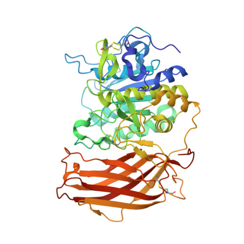
    Inhibitors of the proprotein convertase furin might serve as broad-spectrum antiviral therapeutics. High cellular potency and antiviral activity against acute respiratory syndrome coronavirus 2 (SARS-CoV-2) have been reported for (3,5-dichlorophenyl)pyridine-derived furin inhibitors. Here we characterized the binding mechanism of this inhibitor class using structural, biophysical, and biochemical methods. We established a MALDI-TOF-MS-based furin activity assay, determined IC 50 values, and solved X-ray structures of (3,5-dichlorophenyl)pyridine-derived compounds in complex with furin. The inhibitors induced a substantial conformational rearrangement of the active-site cleft by exposing a central buried tryptophan residue. These changes formed an extended hydrophobic surface patch where the 3,5-dichlorophenyl moiety of the inhibitors was inserted into a newly formed binding pocket. Consistent with these structural rearrangements, we observed slow off-rate binding kinetics and strong structural stabilization in surface plasmon resonance and differential scanning fluorimetry experiments, respectively. The discovered furin conformation offers new opportunities for structure-based drug discovery.


  • Organizational Affiliation
    • Department of Biosciences and Medical Biology, University of Salzburg, Hellbrunner Straße 34, A-5020 Salzburg, Austria.

Macromolecule Content 

  • Total Structure Weight: 53.72 kDa 
  • Atom Count: 4,373 
  • Modeled Residue Count: 471 
  • Deposited Residue Count: 480 
  • Unique protein chains: 1

Macromolecules

Find similar proteins by:|  3D Structure
Entity ID: 1
MoleculeChains  Sequence LengthOrganismDetailsImage
Furin480Homo sapiensMutation(s): 0 
Gene Names: FURINFURPACEPCSK3
EC: 3.4.21.75
UniProt & NIH Common Fund Data Resources
Find proteins for P09958 (Homo sapiens)
Explore P09958 
Go to UniProtKB:  P09958
PHAROS:  P09958
GTEx:  ENSG00000140564 
Entity Groups
Sequence Clusters30% Identity50% Identity70% Identity90% Identity95% Identity100% Identity
UniProt GroupP09958
Glycosylation
Glycosylation Sites: 1Go to GlyGen: P09958-1
Sequence Annotations
Expand
Reference Sequence

Small Molecules

Ligands 6 Unique
IDChains Name / Formula / InChI Key2D Diagram3D Interactions
I0Q
(Subject of Investigation/LOI)

Query on I0Q


Download:Ideal Coordinates CCD File
B [auth A](2R)-4-[4-[5-[4-[[4-(acetamidomethyl)piperidin-1-ium-1-yl]methyl]-6-[3,5-bis(chloranyl)phenyl]pyridin-2-yl]oxypyrimidin-2-yl]piperazin-1-ium-1-yl]-2-methyl-butanoate
C33 H42 Cl2 N7 O4
GOXVJENOKGAJKU-JOCHJYFZSA-O
NAG

Query on NAG


Download:Ideal Coordinates CCD File
Q [auth A]2-acetamido-2-deoxy-beta-D-glucopyranose
C8 H15 N O6
OVRNDRQMDRJTHS-FMDGEEDCSA-N
DMS

Query on DMS


Download:Ideal Coordinates CCD File
K [auth A]
L [auth A]
M [auth A]
N [auth A]
O [auth A]
K [auth A],
L [auth A],
M [auth A],
N [auth A],
O [auth A],
P [auth A]
DIMETHYL SULFOXIDE
C2 H6 O S
IAZDPXIOMUYVGZ-UHFFFAOYSA-N
CA

Query on CA


Download:Ideal Coordinates CCD File
C [auth A],
D [auth A],
E [auth A]
CALCIUM ION
Ca
BHPQYMZQTOCNFJ-UHFFFAOYSA-N
CL

Query on CL


Download:Ideal Coordinates CCD File
J [auth A]CHLORIDE ION
Cl
VEXZGXHMUGYJMC-UHFFFAOYSA-M
NA

Query on NA


Download:Ideal Coordinates CCD File
F [auth A],
G [auth A],
H [auth A],
I [auth A]
SODIUM ION
Na
FKNQFGJONOIPTF-UHFFFAOYSA-N

Experimental Data & Validation

Experimental Data

  • Method: X-RAY DIFFRACTION
  • Resolution: 1.55 Å
  • R-Value Free:  0.188 (Depositor), 0.188 (DCC) 
  • R-Value Work:  0.165 (Depositor), 0.165 (DCC) 
  • R-Value Observed: 0.166 (Depositor) 
Space Group: P 65 2 2
Unit Cell:
Length ( Å )Angle ( ˚ )
a = 131.369α = 90
b = 131.369β = 90
c = 155.336γ = 120
Software Package:
Software NamePurpose
PHENIXphasing
PHENIXrefinement
XDSdata reduction
XDSdata scaling

Structure Validation

View Full Validation Report


Ligand Structure Quality Assessment 


Entry History 

& Funding Information

Deposition Data


Funding OrganizationLocationGrant Number
Austrian Science FundAustriaM 2730

Revision History  (Full details and data files)

  • Version 1.0: 2022-04-20
    Type: Initial release
  • Version 1.1: 2022-04-27
    Changes: Database references
  • Version 1.2: 2024-01-31
    Changes: Data collection, Refinement description
  • Version 1.3: 2024-11-06
    Changes: Structure summary