7QXE | pdb_00007qxe

Recognition of Staphylococcus aureus wall teichoic acid analogue TB87 (compound 3) by Fab4497


Experimental Data Snapshot

  • Method: X-RAY DIFFRACTION
  • Resolution: 1.84 Å
  • R-Value Free: 
    0.218 (Depositor), 0.215 (DCC) 
  • R-Value Work: 
    0.188 (Depositor) 

Starting Model: experimental
View more details

wwPDB Validation 3D Report Full Report

Validation slider image for 7QXE

Ligand Structure Quality Assessment 


This is version 1.3 of the entry. See complete history

Literature

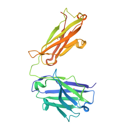
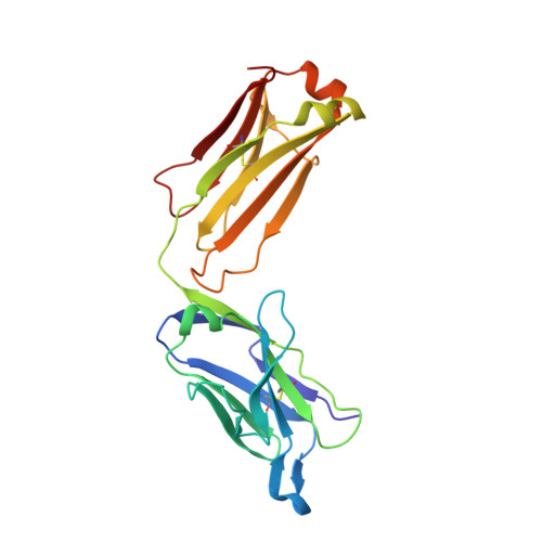
Antibody Recognition of Different Staphylococcus aureus Wall Teichoic Acid Glycoforms.

Di Carluccio, C.Soriano-Maldonado, P.Berni, F.de Haas, C.J.C.Temming, A.R.Hendriks, A.Ali, S.Molinaro, A.Silipo, A.van Sorge, N.M.van Raaij, M.J.Codee, J.D.C.Marchetti, R.

(2022) ACS Cent Sci 8: 1383-1392

  • DOI: https://doi.org/10.1021/acscentsci.2c00125
  • Primary Citation Related Structures: 
    7QXC, 7QXD, 7QXE

  • PubMed Abstract: 

    Wall teichoic acids (WTAs) are glycopolymers decorating the surface of Gram-positive bacteria and potential targets for antibody-mediated treatments against Staphylococcus aureus , including methicillin-resistant (MRSA) strains. Through a combination of glycan microarray, synthetic chemistry, crystallography, NMR, and computational studies, we unraveled the molecular and structural details of fully defined synthetic WTA fragments recognized by previously described monoclonal antibodies (mAbs 4461 and 4497). Our results unveiled the structural requirements for the discriminatory recognition of α- and β-GlcNAc-modified WTA glycoforms by the complementarity-determining regions (CDRs) of the heavy and light chains of the mAbs. Both mAbs interacted not only with the sugar moiety but also with the phosphate groups as well as residues in the ribitol phosphate (RboP) units of the WTA backbone, highlighting their significant role in ligand specificity. Using elongated WTA fragments, containing two sugar modifications, we also demonstrated that the internal carbohydrate moiety of α-GlcNAc-modified WTA is preferentially accommodated in the binding pocket of mAb 4461 with respect to the terminal moiety. Our results also explained the recently documented cross-reactivity of mAb 4497 for β-1,3/β-1,4-GlcNAc-modified WTA, revealing that the flexibility of the RboP backbone is crucial to allow positioning of both glycans in the antibody binding pocket.


  • Organizational Affiliation
    • Department of Chemical Sciences, University of Naples Federico II, Via Cinthia 4, 80126Naples, Italy.

Macromolecule Content 

  • Total Structure Weight: 101.14 kDa 
  • Atom Count: 7,296 
  • Modeled Residue Count: 868 
  • Deposited Residue Count: 912 
  • Unique protein chains: 2

Macromolecules

Find similar proteins by:|  3D Structure
Entity ID: 1
MoleculeChains  Sequence LengthOrganismDetailsImage
Antibody Fab 4497 heavy chainA [auth HHH],
C [auth KKK]
236Homo sapiensMutation(s): 0 
Find similar proteins by:|  3D Structure
Entity ID: 2
MoleculeChains  Sequence LengthOrganismDetailsImage
Antibody 4497 light chainB [auth LLL],
D [auth MMM]
220Homo sapiensMutation(s): 0 

Small Molecules

Ligands 5 Unique
IDChains Name / Formula / InChI Key2D Diagram3D Interactions
G7C
(Subject of Investigation/LOI)

Query on G7C


Download:Ideal Coordinates CCD File
E [auth HHH],
K [auth KKK]
[(2~{S},4~{R})-3-[(2~{S},3~{R},4~{R},5~{S},6~{R})-3-acetamido-6-(hydroxymethyl)-4,5-bis(oxidanyl)oxan-2-yl]oxy-2,4-bis(oxidanyl)-5-[oxidanyl-[(2~{S})-2,3,4,5-tetrakis(oxidanyl)pentoxy]phosphoryl]oxy-pentyl] [(2~{R},3~{S})-2,3,4,5-tetrakis(oxidanyl)pentyl] hydrogen phosphate
C23 H47 N O24 P2
UQOCSXCRLULBKU-APIFBWIWSA-N
PEG

Query on PEG


Download:Ideal Coordinates CCD File
F [auth HHH],
H [auth LLL]
DI(HYDROXYETHYL)ETHER
C4 H10 O3
MTHSVFCYNBDYFN-UHFFFAOYSA-N
SO4

Query on SO4


Download:Ideal Coordinates CCD File
I [auth LLL],
J [auth LLL],
M [auth MMM],
N [auth MMM],
O [auth MMM]
SULFATE ION
O4 S
QAOWNCQODCNURD-UHFFFAOYSA-L
GOL

Query on GOL


Download:Ideal Coordinates CCD File
L [auth KKK]GLYCEROL
C3 H8 O3
PEDCQBHIVMGVHV-UHFFFAOYSA-N
CL

Query on CL


Download:Ideal Coordinates CCD File
G [auth HHH]CHLORIDE ION
Cl
VEXZGXHMUGYJMC-UHFFFAOYSA-M

Experimental Data & Validation

Experimental Data

  • Method: X-RAY DIFFRACTION
  • Resolution: 1.84 Å
  • R-Value Free:  0.218 (Depositor), 0.215 (DCC) 
  • R-Value Work:  0.188 (Depositor) 
Space Group: P 21 21 21
Unit Cell:
Length ( Å )Angle ( ˚ )
a = 64.033α = 90
b = 112.915β = 90
c = 154.993γ = 90
Software Package:
Software NamePurpose
REFMACrefinement
XDSdata reduction
Aimlessdata scaling
REFMACphasing

Structure Validation

View Full Validation Report


Ligand Structure Quality Assessment 


Entry History 

& Funding Information

Deposition Data


Funding OrganizationLocationGrant Number
Ministerio de Ciencia e Innovacion (MCIN)SpainBFU2017-82207-P

Revision History  (Full details and data files)

  • Version 1.0: 2022-08-31
    Type: Initial release
  • Version 1.1: 2022-11-09
    Changes: Database references
  • Version 1.2: 2024-01-31
    Changes: Data collection, Refinement description
  • Version 1.3: 2024-11-06
    Changes: Structure summary