


Structure of M.BseCI DNA methyltransferase from Geobacillus stearothermophilus.
Mitsikas, D.A., Kouyianou, K., Kotsifaki, D., Providaki, M., Bouriotis, V., Glykos, N.M., Kokkinidis, M.To be published.
Experimental Data Snapshot
Entity ID: 1 | |||||
|---|---|---|---|---|---|
| Molecule | Chains | Sequence Length | Organism | Details | Image |
| Modification methylase BseCI | 585 | Geobacillus stearothermophilus | Mutation(s): 0 Gene Names: bseCIM EC: 2.1.1.72 | 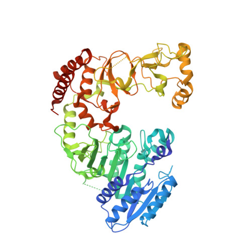 | |
UniProt | |||||
Entity Groups | |||||
| Sequence Clusters | 30% Identity50% Identity70% Identity90% Identity95% Identity100% Identity | ||||
| UniProt Group | P43423 | ||||
Sequence AnnotationsExpand | |||||
Reference Sequence | |||||
Entity ID: 2 | ||||
| Molecule | Chains | Length | Organism | Image |
|---|---|---|---|---|
| Unmethylated DNA duplex | B [auth Z] | 10 | Geobacillus stearothermophilus | 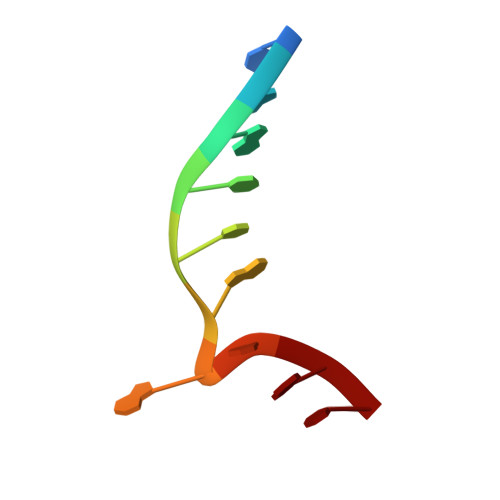 |
Sequence AnnotationsExpand | ||||
Reference Sequence | ||||
Entity ID: 3 | ||||
| Molecule | Chains | Length | Organism | Image |
|---|---|---|---|---|
| Unmethylated DNA duplex | C [auth Y] | 10 | Geobacillus stearothermophilus | 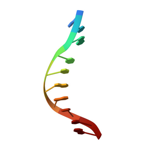 |
Sequence AnnotationsExpand | ||||
Reference Sequence | ||||
| Ligands 1 Unique | |||||
|---|---|---|---|---|---|
| ID | Chains | Name / Formula / InChI Key | 2D Diagram | 3D Interactions | |
| SAH (Subject of Investigation/LOI) Download:Ideal Coordinates CCD File | D [auth A] | S-ADENOSYL-L-HOMOCYSTEINE C14 H20 N6 O5 S ZJUKTBDSGOFHSH-WFMPWKQPSA-N |  | ||
| Length ( Å ) | Angle ( ˚ ) |
|---|---|
| a = 87.04 | α = 90 |
| b = 87.04 | β = 90 |
| c = 156.35 | γ = 120 |
| Software Name | Purpose |
|---|---|
| PHENIX | refinement |
| iMOSFLM | data reduction |
| Aimless | data scaling |
| PHASER | phasing |
| CRANK | phasing |
| BUCCANEER | model building |
| Funding Organization | Location | Grant Number |
|---|---|---|
| Not funded | Greece | -- |


