7QVK | pdb_00007qvk

NM-02 in complex with HER2-ECD


Experimental Data Snapshot

  • Method: X-RAY DIFFRACTION
  • Resolution: 3.10 Å
  • R-Value Free: 
    0.330 (Depositor), 0.338 (DCC) 
  • R-Value Work: 
    0.275 (Depositor), 0.282 (DCC) 
  • R-Value Observed: 
    0.275 (Depositor) 

Starting Model: experimental
View more details

wwPDB Validation 3D Report Full Report

Validation slider image for 7QVK

Ligand Structure Quality Assessment 


This is version 1.3 of the entry. See complete history

Literature

Co-crystallisation and humanisation of an anti-HER2 single-domain antibody as a theranostic tool.

Sawmynaden, K.Wong, N.Davies, S.Cowan, R.Brown, R.Tang, D.Henry, M.Tickle, D.Matthews, D.Carr, M.Bakrania, P.Hoi Ting, H.Hall, G.

(2023) PLoS One 18: e0288259-e0288259

  • DOI: https://doi.org/10.1371/journal.pone.0288259
  • Primary Citation Related Structures: 
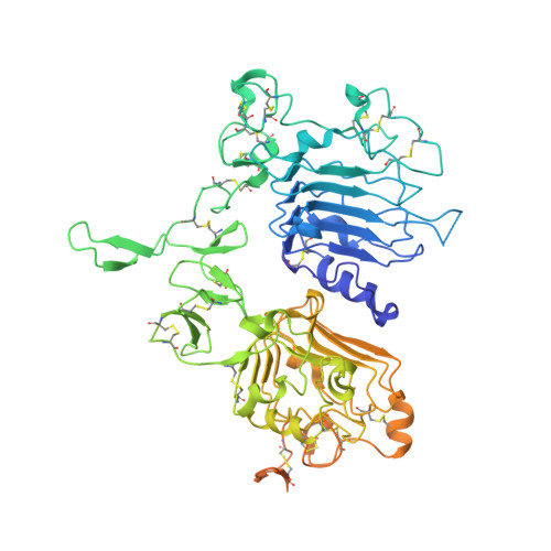
    7QVK

  • PubMed Abstract: 

    Human epidermal growth factor receptor-2 (HER2) is a well-recognised biomarker associated with 25% of breast cancers. In most cases, early detection and/or treatment correlates with an increased chance of survival. This study, has identified and characterised a highly specific anti-HER2 single-domain antibody (sdAb), NM-02, as a potential theranostic tool. Complete structural description by X-ray crystallography has revealed a non-overlapping epitope with current anti-HER2 antibodies. To reduce the immunogenicity risk, NM-02 underwent a humanisation process and retained wild type-like binding properties. To further de-risk the progression towards chemistry, manufacturing and control (CMC) we performed full developability profiling revealing favourable thermal and physical biochemical 'drug-like' properties. Finally, the application of the lead humanised NM-02 candidate (variant K) for HER2-specific imaging purposes was demonstrated using breast cancer HER2+/BT474 xenograft mice.


  • Organizational Affiliation
    • LifeArc, Open Innovation Campus, Stevenage, United Kingdom.

Macromolecule Content 

  • Total Structure Weight: 84.77 kDa 
  • Atom Count: 4,836 
  • Modeled Residue Count: 627 
  • Deposited Residue Count: 762 
  • Unique protein chains: 2

Macromolecules

Find similar proteins by:|  3D Structure
Entity ID: 1
MoleculeChains  Sequence LengthOrganismDetailsImage
Receptor tyrosine-protein kinase erbB-2A [auth AAA]630Homo sapiensMutation(s): 0 
Gene Names: ERBB2HER2MLN19NEUNGL
EC: 2.7.10.1
UniProt & NIH Common Fund Data Resources
Find proteins for P04626 (Homo sapiens)
Explore P04626 
Go to UniProtKB:  P04626
PHAROS:  P04626
GTEx:  ENSG00000141736 
Entity Groups
Sequence Clusters30% Identity50% Identity70% Identity90% Identity95% Identity100% Identity
UniProt GroupP04626
Glycosylation
Glycosylation Sites: 2Go to GlyGen: P04626-1
Sequence Annotations
Expand
Reference Sequence
Find similar proteins by:|  3D Structure
Entity ID: 2
MoleculeChains  Sequence LengthOrganismDetailsImage
NM-02B [auth BBB]132Camelus bactrianusMutation(s): 0 

Oligosaccharides

Help  
Entity ID: 3
MoleculeChains Length2D Diagram GlycosylationD Interactions
beta-D-mannopyranose-(1-4)-2-acetamido-2-deoxy-beta-D-glucopyranose-(1-4)-2-acetamido-2-deoxy-beta-D-glucopyranoseC [auth AbA]3N-Glycosylation
Glycosylation Resources
GlyTouCan: G15407YE
GlyCosmos: G15407YE
GlyGen: G15407YE

Small Molecules

Ligands 1 Unique
IDChains Name / Formula / InChI Key2D Diagram3D Interactions
NAG

Query on NAG


Download:Ideal Coordinates CCD File
D [auth AAA]2-acetamido-2-deoxy-beta-D-glucopyranose
C8 H15 N O6
OVRNDRQMDRJTHS-FMDGEEDCSA-N

Experimental Data & Validation

Experimental Data

  • Method: X-RAY DIFFRACTION
  • Resolution: 3.10 Å
  • R-Value Free:  0.330 (Depositor), 0.338 (DCC) 
  • R-Value Work:  0.275 (Depositor), 0.282 (DCC) 
  • R-Value Observed: 0.275 (Depositor) 
Space Group: P 32 2 1
Unit Cell:
Length ( Å )Angle ( ˚ )
a = 105.767α = 90
b = 105.767β = 90
c = 185.882γ = 120
Software Package:
Software NamePurpose
REFMACrefinement
XDSdata reduction
Aimlessdata scaling
PHASERphasing

Structure Validation

View Full Validation Report


Ligand Structure Quality Assessment 


Entry History 

& Funding Information

Deposition Data


Funding OrganizationLocationGrant Number
Other private--

Revision History  (Full details and data files)

  • Version 1.0: 2023-02-01
    Type: Initial release
  • Version 1.1: 2023-07-26
    Changes: Database references, Derived calculations
  • Version 1.2: 2024-02-07
    Changes: Data collection, Refinement description
  • Version 1.3: 2024-11-06
    Changes: Structure summary