7QU2 | pdb_00007qu2

Junin virus GP1 glycoprotein in complex with Fab fragment of antibody JUN1


Experimental Data Snapshot

  • Method: X-RAY DIFFRACTION
  • Resolution: 2.50 Å
  • R-Value Free: 
    0.254 (Depositor), 0.253 (DCC) 
  • R-Value Work: 
    0.223 (Depositor), 0.222 (DCC) 
  • R-Value Observed: 
    0.225 (Depositor) 

Starting Model: experimental
View more details

wwPDB Validation 3D Report Full Report

Validation slider image for 7QU2

Ligand Structure Quality Assessment 


This is version 1.3 of the entry. See complete history

Literature

Contrasting Modes of New World Arenavirus Neutralization by Immunization-Elicited Monoclonal Antibodies.

Ng, W.M.Sahin, M.Krumm, S.A.Seow, J.Zeltina, A.Harlos, K.Paesen, G.C.Pinschewer, D.D.Doores, K.J.Bowden, T.A.

(2022) mBio 13: e0265021-e0265021

  • DOI: https://doi.org/10.1128/mbio.02650-21
  • Primary Citation Related Structures: 
    7QU1, 7QU2

  • PubMed Abstract: 

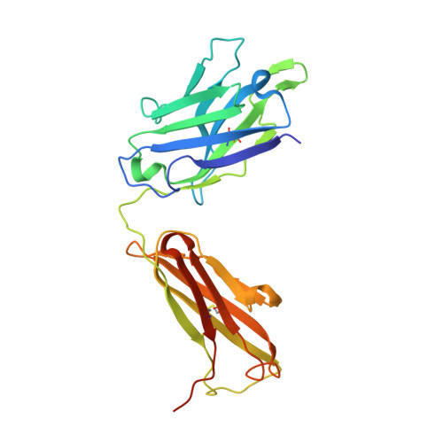
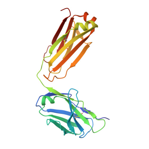
    Transmission of the New World hemorrhagic fever arenaviruses Junín virus (JUNV) and Machupo virus (MACV) to humans is facilitated, in part, by the interaction between the arenavirus GP1 glycoprotein and the human transferrin receptor 1 (hTfR1). We utilize a mouse model of live-attenuated immunization with envelope exchange viruses to isolate neutralizing monoclonal antibodies (NAbs) specific to JUNV GP1 and MACV GP1. Structures of two NAbs, termed JUN1 and MAC1, demonstrate that they neutralize through disruption of hTfR1 recognition. JUN1 utilizes a binding mode common to all characterized infection- and vaccine-elicited JUNV-specific NAbs, which involves mimicking hTfR1 binding through the insertion of a tyrosine into the receptor-binding site. In contrast, MAC1 undergoes a tyrosine-mediated mode of antigen recognition distinct from that used by the reported anti-JUNV NAbs and the only other characterized anti-MACV NAb. These data reveal the varied modes of GP1-specific recognition among New World arenaviruses by the antibody-mediated immune response. IMPORTANCE The GP1 subcomponent of the New World arenavirus GP is a primary target of the neutralizing antibody response, which has been shown to be effective in the prevention and treatment of infection. Here, we characterize the structural basis of the antibody-mediated immune response that arises from immunization of mice against Junín virus and Machupo virus, two rodent-borne zoonotic New World arenaviruses. We isolate a panel of GP1-specific monoclonal antibodies that recognize overlapping epitopes and exhibit neutralizing behavior, in vitro . Structural characterization of two of these antibodies indicates that antibody recognition likely interferes with GP1-mediated recognition of the transferrin receptor 1. These data provide molecular-level detail for a key region of vulnerability on the New World arenavirus surface and a blueprint for therapeutic antibody development.


  • Organizational Affiliation
    • Division of Structural Biology, Wellcome Centre for Human Genetics, University of Oxfordgrid.4991.5, Oxford, United Kingdom.

Macromolecule Content 

  • Total Structure Weight: 69.27 kDa 
  • Atom Count: 4,666 
  • Modeled Residue Count: 586 
  • Deposited Residue Count: 615 
  • Unique protein chains: 3

Macromolecules

Find similar proteins by:|  3D Structure
Entity ID: 1
MoleculeChains  Sequence LengthOrganismDetailsImage
Fab JUN1 heavy chain242Mus musculusMutation(s): 0 
Find similar proteins by:|  3D Structure
Entity ID: 2
MoleculeChains  Sequence LengthOrganismDetailsImage
Fab JUN1 light chain217Mus musculusMutation(s): 0 
Find similar proteins by:|  3D Structure
Entity ID: 3
MoleculeChains  Sequence LengthOrganismDetailsImage
Glycoprotein G1156Mammarenavirus juninenseMutation(s): 0 
Gene Names: GPC
UniProt
Find proteins for C1K9J9 (Junin mammarenavirus)
Explore C1K9J9 
Go to UniProtKB:  C1K9J9
Entity Groups
Sequence Clusters30% Identity50% Identity70% Identity90% Identity95% Identity100% Identity
UniProt GroupC1K9J9
Glycosylation
Glycosylation Sites: 2
Sequence Annotations
Expand
Reference Sequence

Oligosaccharides

Help  
Entity ID: 4
MoleculeChains Length2D Diagram GlycosylationD Interactions
2-acetamido-2-deoxy-beta-D-glucopyranose-(1-4)-2-acetamido-2-deoxy-beta-D-glucopyranose
D
2N-Glycosylation
Glycosylation Resources
GlyTouCan: G42666HT
GlyCosmos: G42666HT
GlyGen: G42666HT

Experimental Data & Validation

Experimental Data

  • Method: X-RAY DIFFRACTION
  • Resolution: 2.50 Å
  • R-Value Free:  0.254 (Depositor), 0.253 (DCC) 
  • R-Value Work:  0.223 (Depositor), 0.222 (DCC) 
  • R-Value Observed: 0.225 (Depositor) 
Space Group: P 1 21 1
Unit Cell:
Length ( Å )Angle ( ˚ )
a = 69.847α = 90
b = 66.752β = 103.5
c = 73.101γ = 90
Software Package:
Software NamePurpose
PHENIXrefinement
PDB_EXTRACTdata extraction
xia2data reduction
xia2data scaling
PHASERphasing
Cootmodel building

Structure Validation

View Full Validation Report


Ligand Structure Quality Assessment 


Entry History 

& Funding Information

Deposition Data


Funding OrganizationLocationGrant Number
Wellcome TrustUnited Kingdom203797/Z/16/Z
Medical Research Council (MRC, United Kingdom)United KingdomMR/L009528/1
Medical Research Council (MRC, United Kingdom)United KingdomMR/N002091/1
Medical Research Council (MRC, United Kingdom)United KingdomMR/K024426/1
Medical Research Council (MRC, United Kingdom)United KingdomMR/S007555/1
Swiss National Science FoundationUnited Kingdom310030_173132/1
Wellcome TrustUnited Kingdom203141/Z/16/Z
Medical Research Council (MRC, United Kingdom)United KingdomMR/V031635/1

Revision History  (Full details and data files)

  • Version 1.0: 2022-02-09
    Type: Initial release
  • Version 1.1: 2022-08-24
    Changes: Database references
  • Version 1.2: 2024-01-31
    Changes: Data collection, Refinement description
  • Version 1.3: 2024-11-20
    Changes: Structure summary