


Structural investigations on the Fe(II)/alpha-ketoglutarate dependent dioxygense PlaO1 from Streptomyces sp. Tu6071
Lukat, P.(2011) Thesis
Experimental Data Snapshot
Starting Model: experimental
View more details
(2011) Thesis
Entity ID: 1 | |||||
|---|---|---|---|---|---|
| Molecule | Chains | Sequence Length | Organism | Details | Image |
| PlaO1 | 297 | Streptomyces sp. Tu6071 | Mutation(s): 0 Gene Names: plaO1, STTU_1451 | 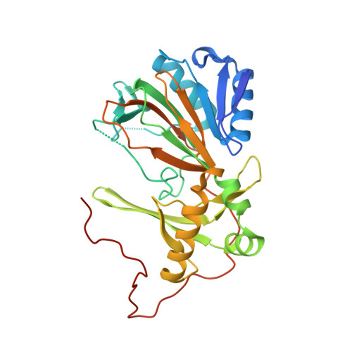 | |
UniProt | |||||
Entity Groups | |||||
| Sequence Clusters | 30% Identity50% Identity70% Identity90% Identity95% Identity100% Identity | ||||
| UniProt Group | Q2I752 | ||||
Sequence AnnotationsExpand | |||||
Reference Sequence | |||||
| Ligands 4 Unique | |||||
|---|---|---|---|---|---|
| ID | Chains | Name / Formula / InChI Key | 2D Diagram | 3D Interactions | |
| SIN (Subject of Investigation/LOI) Download:Ideal Coordinates CCD File | F [auth A], I [auth B], L [auth C], N [auth D] | SUCCINIC ACID C4 H6 O4 KDYFGRWQOYBRFD-UHFFFAOYSA-N |  | ||
| MLI Download:Ideal Coordinates CCD File | E [auth A], K [auth C] | MALONATE ION C3 H2 O4 OFOBLEOULBTSOW-UHFFFAOYSA-L |  | ||
| ACT Download:Ideal Coordinates CCD File | G [auth A] | ACETATE ION C2 H3 O2 QTBSBXVTEAMEQO-UHFFFAOYSA-M |  | ||
| NA Download:Ideal Coordinates CCD File | H [auth A], J [auth B], M [auth C], O [auth D] | SODIUM ION Na FKNQFGJONOIPTF-UHFFFAOYSA-N |  | ||
| Length ( Å ) | Angle ( ˚ ) |
|---|---|
| a = 63.894 | α = 90 |
| b = 108.172 | β = 93.87 |
| c = 115.033 | γ = 90 |
| Software Name | Purpose |
|---|---|
| PHENIX | refinement |
| autoPROC | data reduction |
| Aimless | data scaling |
| MOLREP | phasing |
| PDB_EXTRACT | data extraction |
| Funding Organization | Location | Grant Number |
|---|---|---|
| Not funded | -- |


