7QRW | pdb_00007qrw

Crystal structure of NHL domain of TRIM3


Experimental Data Snapshot

  • Method: X-RAY DIFFRACTION
  • Resolution: 1.70 Å
  • R-Value Free: 
    0.215 (Depositor), 0.218 (DCC) 
  • R-Value Work: 
    0.182 (Depositor), 0.192 (DCC) 
  • R-Value Observed: 
    0.184 (Depositor) 

Starting Model: experimental
View more details

wwPDB Validation 3D Report Full Report

Validation slider image for 7QRW

This is version 1.2 of the entry. See complete history

Literature

Comparative structural analyses of the NHL domains from the human E3 ligase TRIM-NHL family.

Chaikuad, A.Zhubi, R.Tredup, C.Knapp, S.

(2022) IUCrJ 9: 720-727

  • DOI: https://doi.org/10.1107/S2052252522008582
  • Primary Citation Related Structures: 
    7QRV, 7QRW, 7QRX

  • PubMed Abstract: 

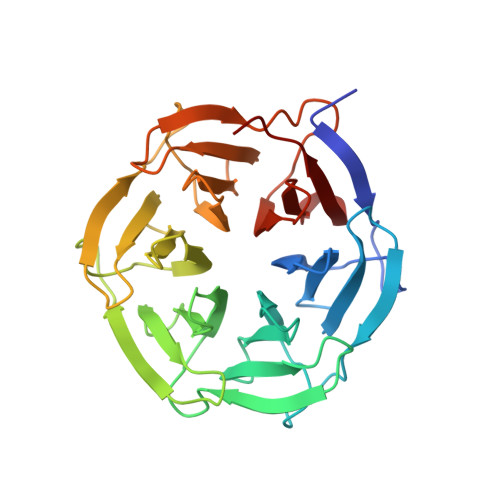
    Tripartite motif (TRIM) proteins constitute one of the largest subfamilies of the RING-type E3 ubiquitin ligases that play a role in diverse processes from homeostasis and immune response to viral restriction. While TRIM proteins typically harbor an N-terminal RING finger, a B-box and a coiled-coil domain, a high degree of diversity lies in their C termini that contain diverse protein interaction modules, most of which, both structures and their roles in intermolecular interactions, remain unknown. Here, high-resolution crystal structures of the NHL domains of three of the four human TRIM-NHL proteins, namely TRIM2, TRIM3 and TRIM71, are presented. Comparative structural analyses revealed that, despite sharing an evolutionarily conserved six-bladed β-propeller architecture, the low sequence identities resulted in distinct properties of these interaction domains at their putative binding sites for macromolecules. Interestingly, residues lining the binding cavities represent a hotspot for genetic mutations linked to several diseases. Thus, high sequence diversity within the conserved NHL domains might be essential for differentiating binding partners among TRIM-NHL proteins.


  • Organizational Affiliation
    • Institute for Pharmaceutical Chemistry, Johann Wolfgang Goethe-University, Max-von-Laue-Strasse 9, D-60438 Frankfurt am Main, Germany.

Macromolecule Content 

  • Total Structure Weight: 30.73 kDa 
  • Atom Count: 2,314 
  • Modeled Residue Count: 276 
  • Deposited Residue Count: 281 
  • Unique protein chains: 1

Macromolecules

Find similar proteins by:|  3D Structure
Entity ID: 1
MoleculeChains  Sequence LengthOrganismDetailsImage
Isoform 4 of Tripartite motif-containing protein 3281Homo sapiensMutation(s): 0 
Gene Names: TRIM3BERPRNF22RNF97
EC: 2.3.2.27
UniProt & NIH Common Fund Data Resources
Find proteins for O75382 (Homo sapiens)
Explore O75382 
Go to UniProtKB:  O75382
PHAROS:  O75382
GTEx:  ENSG00000110171 
Entity Groups
Sequence Clusters30% Identity50% Identity70% Identity90% Identity95% Identity100% Identity
UniProt GroupO75382
Sequence Annotations
Expand
Reference Sequence

Experimental Data & Validation

Experimental Data

  • Method: X-RAY DIFFRACTION
  • Resolution: 1.70 Å
  • R-Value Free:  0.215 (Depositor), 0.218 (DCC) 
  • R-Value Work:  0.182 (Depositor), 0.192 (DCC) 
  • R-Value Observed: 0.184 (Depositor) 
Space Group: P 41 21 2
Unit Cell:
Length ( Å )Angle ( ˚ )
a = 58.933α = 90
b = 58.933β = 90
c = 161.229γ = 90
Software Package:
Software NamePurpose
Aimlessdata scaling
REFMACrefinement
PDB_EXTRACTdata extraction
XDSdata reduction
PHASERphasing

Structure Validation

View Full Validation Report


Entry History 

& Funding Information

Deposition Data


Funding OrganizationLocationGrant Number
Not funded--

Revision History  (Full details and data files)

  • Version 1.0: 2022-05-04
    Type: Initial release
  • Version 1.1: 2022-12-14
    Changes: Database references
  • Version 1.2: 2024-01-31
    Changes: Data collection, Refinement description