Crystal Structures of Botulinum Neurotoxin Subtypes A4 and A5 Cell Binding Domains in Complex with Receptor Ganglioside.
Gregory, K.S., Mojanaga, O.O., Liu, S.M., Acharya, K.R.(2022) Toxins (Basel) 14
- PubMed: 35202156 Search on PubMedSearch on PubMed Central
- DOI: https://doi.org/10.3390/toxins14020129
- Primary Citation Related Structures:
7QPT, 7QPU - PubMed Abstract:
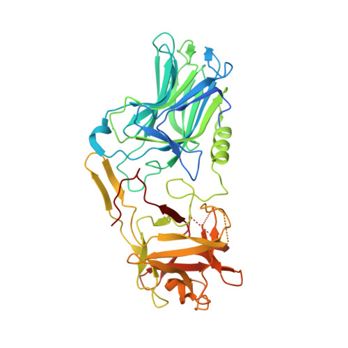
Botulinum neurotoxins (BoNT) cause the potentially fatal neuroparalytic disease botulism that arises due to proteolysis of a SNARE protein. Each BoNT is comprised of three domains: a cell binding domain (H C ), a translocation domain (H N ), and a catalytic (Zn 2+ endopeptidase) domain (LC). The H C is responsible for neuronal specificity by targeting both a protein and ganglioside receptor at the neuromuscular junction. Although highly toxic, some BoNTs are commercially available as therapeutics for the treatment of a range of neuromuscular conditions. Here we present the crystal structures of two BoNT cell binding domains, H C /A4 and H C /A5, in a complex with the oligosaccharide of ganglioside, GD1a and GM1b, respectively. These structures, along with a detailed comparison with the previously reported apo-structures, reveal the conformational changes that occur upon ganglioside binding and the interactions involved.
- Department of Biology and Biochemistry, University of Bath, Claverton Down, Bath BA2 7AY, UK.
Organizational Affiliation: