


Crystal structure of NAD-bound glycosomal malate dehydrogenase from Trypanosoma cruzi
Sonani, R.R., Dubin, G.To be published.
Experimental Data Snapshot
Starting Model: experimental
View more details
Entity ID: 1 | |||||
|---|---|---|---|---|---|
| Molecule | Chains | Sequence Length | Organism | Details | Image |
| Malate dehydrogenase | 331 | Trypanosoma cruzi | Mutation(s): 0 Gene Names: C3747_2g261, ECC02_003170 EC: 1.1.1.37 | 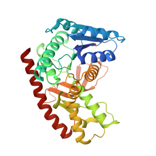 | |
UniProt | |||||
Entity Groups | |||||
| Sequence Clusters | 30% Identity50% Identity70% Identity90% Identity95% Identity100% Identity | ||||
| UniProt Group | A0A2V2XLH9 | ||||
Sequence AnnotationsExpand | |||||
Reference Sequence | |||||
| Ligands 2 Unique | |||||
|---|---|---|---|---|---|
| ID | Chains | Name / Formula / InChI Key | 2D Diagram | 3D Interactions | |
| NAD Download:Ideal Coordinates CCD File | I [auth A] L [auth B] N [auth C] R [auth D] S [auth E] | NICOTINAMIDE-ADENINE-DINUCLEOTIDE C21 H27 N7 O14 P2 BAWFJGJZGIEFAR-NNYOXOHSSA-N |  | ||
| GOL (Subject of Investigation/LOI) Download:Ideal Coordinates CCD File | J [auth A] K [auth A] M [auth B] O [auth C] P [auth C] | GLYCEROL C3 H8 O3 PEDCQBHIVMGVHV-UHFFFAOYSA-N |  | ||
| Length ( Å ) | Angle ( ˚ ) |
|---|---|
| a = 80.481 | α = 97.6 |
| b = 84.802 | β = 100.44 |
| c = 113.459 | γ = 110.08 |
| Software Name | Purpose |
|---|---|
| REFMAC | refinement |
| PDB_EXTRACT | data extraction |
| XDS | data reduction |
| Aimless | data scaling |
| PHASER | phasing |
| Funding Organization | Location | Grant Number |
|---|---|---|
| Polish National Science Centre | Poland | 2017/26/M/NZ1/00797 |
| Foundation for Polish Science | Poland | TEAM TECH CORE FACILITY/2017-4/6 |


