


Crystal structure of Mycobacterium hassiacum glucosyl-3-phosphoglycerate synthase at pH 8.5 in complex with UMP and Magnesium
Silva, A., Nunes-Costa, D., Empadinhas, N., Barbosa Pereira, P.J., Macedo-Ribeiro, S.To be published.
Experimental Data Snapshot
Starting Model: experimental
View more details
Entity ID: 1 | |||||
|---|---|---|---|---|---|
| Molecule | Chains | Sequence Length | Organism | Details | Image |
| Glucosyl-3-phosphoglycerate synthase | 327 | Mycolicibacterium hassiacum DSM 44199 | Mutation(s): 0 Gene Names: gpgS, C731_3243, MHAS_02845 EC: 2.4.1.266 | 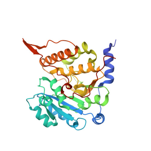 | |
UniProt | |||||
Entity Groups | |||||
| Sequence Clusters | 30% Identity50% Identity70% Identity90% Identity95% Identity100% Identity | ||||
| UniProt Group | K5B7Z4 | ||||
Sequence AnnotationsExpand | |||||
Reference Sequence | |||||
| Ligands 3 Unique | |||||
|---|---|---|---|---|---|
| ID | Chains | Name / Formula / InChI Key | 2D Diagram | 3D Interactions | |
| U5P (Subject of Investigation/LOI) Download:Ideal Coordinates CCD File | D [auth A], H [auth B] | URIDINE-5'-MONOPHOSPHATE C9 H13 N2 O9 P DJJCXFVJDGTHFX-XVFCMESISA-N |  | ||
| CL Download:Ideal Coordinates CCD File | E [auth A] | CHLORIDE ION Cl VEXZGXHMUGYJMC-UHFFFAOYSA-M |  | ||
| MG Download:Ideal Coordinates CCD File | C [auth A], F [auth A], G [auth B], I [auth B] | MAGNESIUM ION Mg JLVVSXFLKOJNIY-UHFFFAOYSA-N |  | ||
| Length ( Å ) | Angle ( ˚ ) |
|---|---|
| a = 71.076 | α = 90 |
| b = 90.377 | β = 90 |
| c = 96.007 | γ = 90 |
| Software Name | Purpose |
|---|---|
| PHENIX | refinement |
| XDS | data reduction |
| SCALA | data scaling |
| PHASER | phasing |
| Funding Organization | Location | Grant Number |
|---|---|---|
| Fundacao para a Ciencia e a Tecnologia | Portugal | PTDC/BTM-TEC/29221/2017 (POCI-01-0145-FEDER-029221). |


