7QOF | pdb_00007qof

Icosahedral capsid of the phicrAss001 virion


Experimental Data Snapshot

  • Method: ELECTRON MICROSCOPY
  • Resolution: 3.01 Å
  • Aggregation State: PARTICLE 
  • Reconstruction Method: SINGLE PARTICLE 

wwPDB Validation 3D Report Full Report

Validation slider image for 7QOF

This is version 1.4 of the entry. See complete history

Literature

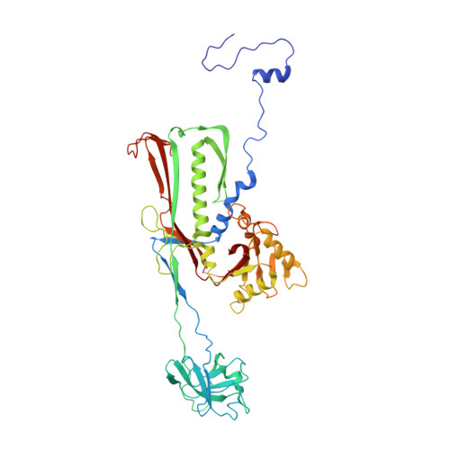
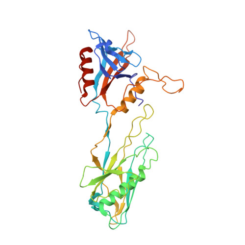
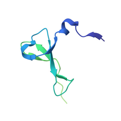
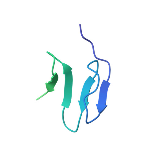
Structural atlas of a human gut crassvirus.

Bayfield, O.W.Shkoporov, A.N.Yutin, N.Khokhlova, E.V.Smith, J.L.R.Hawkins, D.E.D.P.Koonin, E.V.Hill, C.Antson, A.A.

(2023) Nature 617: 409-416

  • DOI: https://doi.org/10.1038/s41586-023-06019-2
  • Primary Citation Related Structures: 
    7QOF, 7QOG, 7QOH, 7QOI, 7QOJ, 7QOK, 7QOL

  • PubMed Abstract: 

    CrAssphage and related viruses of the order Crassvirales (hereafter referred to as crassviruses) were originally discovered by cross-assembly of metagenomic sequences. They are the most abundant viruses in the human gut, are found in the majority of individual gut viromes, and account for up to 95% of the viral sequences in some individuals 1-4 . Crassviruses are likely to have major roles in shaping the composition and functionality of the human microbiome, but the structures and roles of most of the virally encoded proteins are unknown, with only generic predictions resulting from bioinformatic analyses 4,5 . Here we present a cryo-electron microscopy reconstruction of Bacteroides intestinalis virus ΦcrAss001 6 , providing the structural basis for the functional assignment of most of its virion proteins. The muzzle protein forms an assembly about 1 MDa in size at the end of the tail and exhibits a previously unknown fold that we designate the 'crass fold', that is likely to serve as a gatekeeper that controls the ejection of cargos. In addition to packing the approximately 103 kb of virus DNA, the ΦcrAss001 virion has extensive storage space for virally encoded cargo proteins in the capsid and, unusually, within the tail. One of the cargo proteins is present in both the capsid and the tail, suggesting a general mechanism for protein ejection, which involves partial unfolding of proteins during their extrusion through the tail. These findings provide a structural basis for understanding the mechanisms of assembly and infection of these highly abundant crassviruses.


  • Organizational Affiliation
    • York Structural Biology Laboratory, Department of Chemistry, University of York, York, UK. oliver.bayfield@york.ac.uk.

Macromolecule Content 

  • Total Structure Weight: 875.82 kDa 
  • Atom Count: 59,983 
  • Modeled Residue Count: 7,647 
  • Deposited Residue Count: 7,874 
  • Unique protein chains: 4

Macromolecules

Find similar proteins by:|  3D Structure
Entity ID: 1
MoleculeChains  Sequence LengthOrganismDetailsImage
Major capsid protein gp32
A, B, C, D, E
A, B, C, D, E, F, G, H, I
504Bacteroides phage crAss001Mutation(s): 0 
UniProt
Find proteins for A0A385DVU6 (Bacteroides phage crAss001)
Explore A0A385DVU6 
Go to UniProtKB:  A0A385DVU6
Entity Groups
Sequence Clusters30% Identity50% Identity70% Identity90% Identity95% Identity100% Identity
UniProt GroupA0A385DVU6
Sequence Annotations
Expand
Reference Sequence
Find similar proteins by:|  3D Structure
Entity ID: 2
MoleculeChains  Sequence LengthOrganismDetailsImage
Auxiliary capsid protein gp36333Bacteroides phage crAss001Mutation(s): 0 
UniProt
Find proteins for A0A385DVS7 (Bacteroides phage crAss001)
Explore A0A385DVS7 
Go to UniProtKB:  A0A385DVS7
Entity Groups
Sequence Clusters30% Identity50% Identity70% Identity90% Identity95% Identity100% Identity
UniProt GroupA0A385DVS7
Sequence Annotations
Expand
Reference Sequence
Find similar proteins by:|  3D Structure
Entity ID: 3
MoleculeChains  Sequence LengthOrganismDetailsImage
Head fiber trimer protein gp21S [auth j],
T [auth k]
122Bacteroides phage crAss001Mutation(s): 0 
UniProt
Find proteins for A0A385DVL5 (Bacteroides phage crAss001)
Explore A0A385DVL5 
Go to UniProtKB:  A0A385DVL5
Entity Groups
Sequence Clusters30% Identity50% Identity70% Identity90% Identity95% Identity100% Identity
UniProt GroupA0A385DVL5
Sequence Annotations
Expand
Reference Sequence
Find similar proteins by:|  3D Structure
Entity ID: 4
MoleculeChains  Sequence LengthOrganismDetailsImage
Head fiber dimer protein gp29U [auth l]97Bacteroides phage crAss001Mutation(s): 0 
UniProt
Find proteins for A0A385DTC5 (Bacteroides phage crAss001)
Explore A0A385DTC5 
Go to UniProtKB:  A0A385DTC5
Entity Groups
Sequence Clusters30% Identity50% Identity70% Identity90% Identity95% Identity100% Identity
UniProt GroupA0A385DTC5
Sequence Annotations
Expand
Reference Sequence

Small Molecules

Ligands 1 Unique
IDChains Name / Formula / InChI Key2D Diagram3D Interactions
MG

Query on MG


Download:Ideal Coordinates CCD File
V [auth D]MAGNESIUM ION
Mg
JLVVSXFLKOJNIY-UHFFFAOYSA-N

Experimental Data & Validation

Experimental Data

  • Method: ELECTRON MICROSCOPY
  • Resolution: 3.01 Å
  • Aggregation State: PARTICLE 
  • Reconstruction Method: SINGLE PARTICLE 
EM Software:
TaskSoftware PackageVersion
RECONSTRUCTIONRELION3.1
MODEL REFINEMENTPHENIX

Structure Validation

View Full Validation Report


Entry History 

& Funding Information

Deposition Data


Funding OrganizationLocationGrant Number
Wellcome TrustUnited Kingdom--

Revision History  (Full details and data files)

  • Version 1.0: 2023-03-29
    Type: Initial release
  • Version 1.1: 2023-05-03
    Changes: Database references
  • Version 1.2: 2023-05-17
    Changes: Database references
  • Version 1.3: 2023-05-24
    Changes: Database references
  • Version 1.4: 2024-07-17
    Changes: Data collection