7QMO | pdb_00007qmo

Endothiapepsin in complex with compound TL00150 at room-temperature (temperature ramping down structure 16)


Experimental Data Snapshot

  • Method: X-RAY DIFFRACTION
  • Resolution: 1.79 Å
  • R-Value Free: 
    0.221 (Depositor), 0.221 (DCC) 
  • R-Value Work: 
    0.195 (Depositor), 0.195 (DCC) 
  • R-Value Observed: 
    0.196 (Depositor) 

Starting Model: experimental
View more details

wwPDB Validation 3D Report Full Report

Validation slider image for 7QMO

Ligand Structure Quality Assessment 


This is version 1.2 of the entry. See complete history

Literature

Probing ligand binding of endothiapepsin by `temperature-resolved' macromolecular crystallography.

Huang, C.Y.Aumonier, S.Engilberge, S.Eris, D.Smith, K.M.L.Leonarski, F.Wojdyla, J.A.Beale, J.H.Buntschu, D.Pauluhn, A.Sharpe, M.E.Metz, A.Olieric, V.Wang, M.

(2022) Acta Crystallogr D Struct Biol 78: 964-974

  • DOI: https://doi.org/10.1107/S205979832200612X
  • Primary Citation Related Structures: 
    7QLT, 7QLU, 7QLV, 7QLW, 7QLX, 7QLY, 7QLZ, 7QM0, 7QM1, 7QM3, 7QM4, 7QM5, 7QM6, 7QM7, 7QM8, 7QM9, 7QMA, 7QMB, 7QMC, 7QMD, 7QME, 7QMF, 7QMG, 7QMH, 7QMI, 7QMJ, 7QMK, 7QML, 7QMM, 7QMN, 7QMO, 7QMP, 7QMQ, 7QMR, 7QMS, 7QMT, 7QMU, 7QMV, 7QMW, 7QMX, 7QMY, 7QMZ, 7QN0, 7QN1, 7QN2, 7QN3, 7QN4

  • PubMed Abstract: 

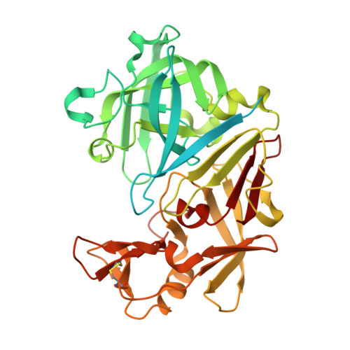
    Continuous developments in cryogenic X-ray crystallography have provided most of our knowledge of 3D protein structures, which has recently been further augmented by revolutionary advances in cryoEM. However, a single structural conformation identified at cryogenic temperatures may introduce a fictitious structure as a result of cryogenic cooling artefacts, limiting the overview of inherent protein physiological dynamics, which play a critical role in the biological functions of proteins. Here, a room-temperature X-ray crystallographic method using temperature as a trigger to record movie-like structural snapshots has been developed. The method has been used to show how TL00150, a 175.15 Da fragment, undergoes binding-mode changes in endothiapepsin. A surprising fragment-binding discrepancy was observed between the cryo-cooled and physiological temperature structures, and multiple binding poses and their interplay with DMSO were captured. The observations here open up new promising prospects for structure determination and interpretation at physiological temperatures with implications for structure-based drug discovery.


  • Organizational Affiliation
    • Swiss Light Source, Paul Scherrer Institut, Forschungsstrasse 111, Villigen-PSI, 5232 Villigen, Switzerland.

Macromolecule Content 

  • Total Structure Weight: 44.04 kDa 
  • Atom Count: 2,631 
  • Modeled Residue Count: 330 
  • Deposited Residue Count: 419 
  • Unique protein chains: 1

Macromolecules

Find similar proteins by:|  3D Structure
Entity ID: 1
MoleculeChains  Sequence LengthOrganismDetailsImage
Endothiapepsin419Cryphonectria parasiticaMutation(s): 0 
EC: 3.4.23.22
UniProt
Find proteins for P11838 (Cryphonectria parasitica)
Explore P11838 
Go to UniProtKB:  P11838
Entity Groups
Sequence Clusters30% Identity50% Identity70% Identity90% Identity95% Identity100% Identity
UniProt GroupP11838
Sequence Annotations
Expand
Reference Sequence

Small Molecules

Ligands 3 Unique
IDChains Name / Formula / InChI Key2D Diagram3D Interactions
PG5

Query on PG5


Download:Ideal Coordinates CCD File
C [auth A]1-METHOXY-2-[2-(2-METHOXY-ETHOXY]-ETHANE
C8 H18 O4
YFNKIDBQEZZDLK-UHFFFAOYSA-N
U1H
(Subject of Investigation/LOI)

Query on U1H


Download:Ideal Coordinates CCD File
B [auth A],
G [auth A]
[4-(trifluoromethyl)phenyl]methanamine
C8 H8 F3 N
PRDBLLIPPDOICK-UHFFFAOYSA-N
DMS

Query on DMS


Download:Ideal Coordinates CCD File
D [auth A],
E [auth A],
F [auth A]
DIMETHYL SULFOXIDE
C2 H6 O S
IAZDPXIOMUYVGZ-UHFFFAOYSA-N

Experimental Data & Validation

Experimental Data

  • Method: X-RAY DIFFRACTION
  • Resolution: 1.79 Å
  • R-Value Free:  0.221 (Depositor), 0.221 (DCC) 
  • R-Value Work:  0.195 (Depositor), 0.195 (DCC) 
  • R-Value Observed: 0.196 (Depositor) 
Space Group: P 1 21 1
Unit Cell:
Length ( Å )Angle ( ˚ )
a = 45.731α = 90
b = 73.906β = 109.72
c = 53.387γ = 90
Software Package:
Software NamePurpose
XDSdata reduction
XSCALEdata scaling
PHASERphasing
PHENIXrefinement

Structure Validation

View Full Validation Report


Ligand Structure Quality Assessment 


Entry History 

& Funding Information

Deposition Data


Funding OrganizationLocationGrant Number
Swiss National Science FoundationEuropean Union182369
Swiss National Science FoundationEuropean Union198290
H2020 Marie Curie Actions of the European CommissionEuropean Union884104

Revision History  (Full details and data files)

  • Version 1.0: 2022-08-17
    Type: Initial release
  • Version 1.1: 2024-01-31
    Changes: Data collection, Refinement description
  • Version 1.2: 2024-10-23
    Changes: Structure summary