7QD7 | pdb_00007qd7

TarM(Se)_G117R


Experimental Data Snapshot

  • Method: X-RAY DIFFRACTION
  • Resolution: 2.06 Å
  • R-Value Free: 
    0.236 (Depositor), 0.240 (DCC) 
  • R-Value Work: 
    0.218 (Depositor), 0.221 (DCC) 

wwPDB Validation 3D Report Full Report

Validation slider image for 7QD7

This is version 1.1 of the entry. See complete history

Literature

Invasive Staphylococcus epidermidis uses a unique processive wall teichoic acid glycosyltransferase to evade immune recognition.

Guo, Y.Du, X.Krusche, J.Beck, C.Ali, S.Walter, A.Winstel, V.Mayer, C.Codee, J.D.C.Peschel, A.Stehle, T.

(2023) Sci Adv 9: eadj2641-eadj2641

  • DOI: https://doi.org/10.1126/sciadv.adj2641
  • Primary Citation Related Structures: 
    7QD7, 7QH9, 7QNT, 8P1X, 8P20

  • PubMed Abstract: 

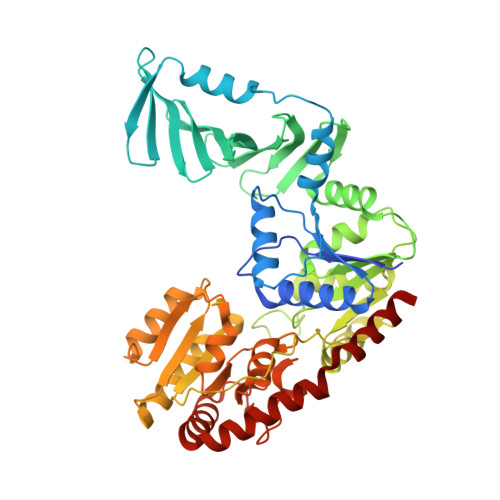
    Staphylococcus epidermidis expresses glycerol phosphate wall teichoic acid (WTA), but some health care-associated methicillin-resistant S. epidermidis (HA-MRSE) clones produce a second, ribitol phosphate (RboP) WTA, resembling that of the aggressive pathogen Staphylococcus aureus . RboP-WTA promotes HA-MRSE persistence and virulence in bloodstream infections. We report here that the TarM enzyme of HA-MRSE [TarM(Se)] glycosylates RboP-WTA with glucose, instead of N -acetylglucosamine (GlcNAc) by TarM(Sa) in S. aureus . Replacement of GlcNAc with glucose in RboP-WTA impairs HA-MRSE detection by human immunoglobulin G, which may contribute to the immune-evasion capacities of many invasive S. epidermidis . Crystal structures of complexes with uridine diphosphate glucose (UDP-glucose), and with UDP and glycosylated poly(RboP), reveal the binding mode and glycosylation mechanism of this enzyme and explain why TarM(Se) and TarM(Sa) link different sugars to poly(RboP). These structural data provide evidence that TarM(Se) is a processive WTA glycosyltransferase. Our study will support the targeted inhibition of TarM enzymes, and the development of RboP-WTA targeting vaccines and phage therapies.


  • Organizational Affiliation
    • Interfaculty Institute of Biochemistry, University of Tübingen, Tübingen, Germany.

Macromolecule Content 

  • Total Structure Weight: 60.46 kDa 
  • Atom Count: 3,983 
  • Modeled Residue Count: 492 
  • Deposited Residue Count: 508 
  • Unique protein chains: 1

Macromolecules

Find similar proteins by:|  3D Structure
Entity ID: 1
MoleculeChains  Sequence LengthOrganismDetailsImage
TarM(Se)_G117RA [auth AAA]508Escherichia coli BL21(DE3)Mutation(s): 0 

Small Molecules

Ligands 4 Unique
IDChains Name / Formula / InChI Key2D Diagram3D Interactions
1PE

Query on 1PE


Download:Ideal Coordinates CCD File
H [auth AAA],
I [auth AAA]
PENTAETHYLENE GLYCOL
C10 H22 O6
JLFNLZLINWHATN-UHFFFAOYSA-N
PO4

Query on PO4


Download:Ideal Coordinates CCD File
F [auth AAA]PHOSPHATE ION
O4 P
NBIIXXVUZAFLBC-UHFFFAOYSA-K
GOL

Query on GOL


Download:Ideal Coordinates CCD File
G [auth AAA]GLYCEROL
C3 H8 O3
PEDCQBHIVMGVHV-UHFFFAOYSA-N
CL

Query on CL


Download:Ideal Coordinates CCD File
B [auth AAA],
C [auth AAA],
D [auth AAA],
E [auth AAA]
CHLORIDE ION
Cl
VEXZGXHMUGYJMC-UHFFFAOYSA-M

Experimental Data & Validation

Experimental Data

  • Method: X-RAY DIFFRACTION
  • Resolution: 2.06 Å
  • R-Value Free:  0.236 (Depositor), 0.240 (DCC) 
  • R-Value Work:  0.218 (Depositor), 0.221 (DCC) 
Space Group: P 21 21 21
Unit Cell:
Length ( Å )Angle ( ˚ )
a = 58.68α = 90
b = 88.42β = 90
c = 97.49γ = 90
Software Package:
Software NamePurpose
REFMACrefinement
XDSdata reduction
XSCALEdata scaling
PHASERphasing

Structure Validation

View Full Validation Report


Entry History 

& Funding Information

Deposition Data


Funding OrganizationLocationGrant Number
German Research Foundation (DFG)GermanyCRC-TR261

Revision History  (Full details and data files)

  • Version 1.0: 2023-05-10
    Type: Initial release
  • Version 1.1: 2023-12-06
    Changes: Data collection, Database references