7QCJ | pdb_00007qcj

Structure of SARS-CoV-2 Papain-like Protease bound to N-(2,4-dihydroxybenzylidene)-thiosemicarbazone


Experimental Data Snapshot

  • Method: X-RAY DIFFRACTION
  • Resolution: 1.84 Å
  • R-Value Free: 
    0.198 (Depositor), 0.200 (DCC) 
  • R-Value Work: 
    0.176 (Depositor), 0.178 (DCC) 
  • R-Value Observed: 
    0.177 (Depositor) 

Starting Model: experimental
View more details

wwPDB Validation 3D Report Full Report

Validation slider image for 7QCJ

Ligand Structure Quality Assessment 


This is version 2.2 of the entry. See complete history

Literature

Hydrazones and Thiosemicarbazones Targeting Protein-Protein-Interactions of SARS-CoV-2 Papain-like Protease.

Ewert, W.Gunther, S.Miglioli, F.Falke, S.Reinke, P.Y.A.Niebling, S.Gunther, C.Han, H.Srinivasan, V.Brognaro, H.Lieske, J.Lorenzen, K.Garcia-Alai, M.M.Betzel, C.Carcelli, M.Hinrichs, W.Rogolino, D.Meents, A.

(2022) Front Chem 10: 832431-832431

  • DOI: https://doi.org/10.3389/fchem.2022.832431
  • Primary Citation Related Structures: 
    7QCG, 7QCH, 7QCI, 7QCJ, 7QCK, 7QCM

  • PubMed Abstract: 

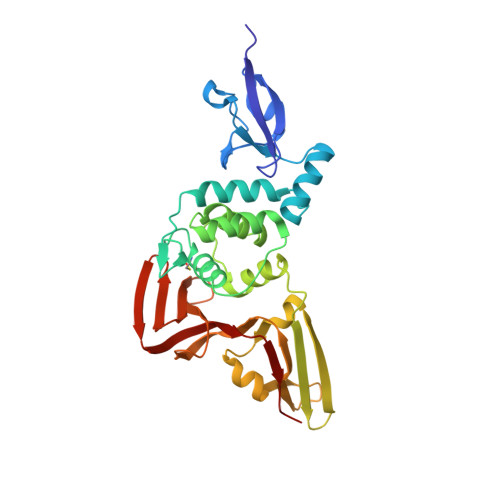
    The papain-like protease (PLpro) of SARS-CoV-2 is essential for viral propagation and, additionally, dysregulation of the host innate immune system. Using a library of 40 potential metal-chelating compounds we performed an X-ray crystallographic screening against PLpro. As outcome we identified six compounds binding to the target protein. Here we describe the interaction of one hydrazone (H1) and five thiosemicarbazone (T1-T5) compounds with the two distinct natural substrate binding sites of PLpro for ubiquitin and ISG15. H1 binds to a polar groove at the S1 binding site by forming several hydrogen bonds with PLpro. T1-T5 bind into a deep pocket close to the polyubiquitin and ISG15 binding site S2. Their interactions are mainly mediated by multiple hydrogen bonds and further hydrophobic interactions. In particular compound H1 interferes with natural substrate binding by sterical hindrance and induces conformational changes in protein residues involved in substrate binding, while compounds T1-T5 could have a more indirect effect. Fluorescence based enzyme activity assay and complementary thermal stability analysis reveal only weak inhibition properties in the high micromolar range thereby indicating the need for compound optimization. Nevertheless, the unique binding properties involving strong hydrogen bonding and the various options for structural optimization make the compounds ideal lead structures. In combination with the inexpensive and undemanding synthesis, the reported hydrazone and thiosemicarbazones represent an attractive scaffold for further structure-based development of novel PLpro inhibitors by interrupting protein-protein interactions at the S1 and S2 site.


  • Organizational Affiliation
    • Center for Free-Electron Laser Science CFEL, Deutsches Elektronen-Synchrotron DESY, Hamburg, Germany.

Macromolecule Content 

  • Total Structure Weight: 36.89 kDa 
  • Atom Count: 2,853 
  • Modeled Residue Count: 315 
  • Deposited Residue Count: 315 
  • Unique protein chains: 1

Macromolecules

Find similar proteins by:|  3D Structure
Entity ID: 1
MoleculeChains  Sequence LengthOrganismDetailsImage
Papain-like protease nsp3315Severe acute respiratory syndrome coronavirus 2Mutation(s): 0 
EC: 3.4.19.12 (PDB Primary Data), 3.4.22 (PDB Primary Data)
UniProt
Find proteins for P0DTD1 (Severe acute respiratory syndrome coronavirus 2)
Explore P0DTD1 
Go to UniProtKB:  P0DTD1
Entity Groups
Sequence Clusters30% Identity50% Identity70% Identity90% Identity95% Identity100% Identity
UniProt GroupP0DTD1
Sequence Annotations
Expand
Reference Sequence

Small Molecules

Ligands 5 Unique
IDChains Name / Formula / InChI Key2D Diagram3D Interactions
A4O
(Subject of Investigation/LOI)

Query on A4O


Download:Ideal Coordinates CCD File
F [auth A]N-(2,4-dihydroxybenzylidene)-thiosemicarbazone
C8 H9 N3 O2 S
DZTQPWJXTVXKJK-ONNFQVAWSA-N
PO4

Query on PO4


Download:Ideal Coordinates CCD File
I [auth A],
J [auth A],
K [auth A]
PHOSPHATE ION
O4 P
NBIIXXVUZAFLBC-UHFFFAOYSA-K
GOL

Query on GOL


Download:Ideal Coordinates CCD File
B [auth A],
C [auth A],
D [auth A],
E [auth A],
G [auth A]
GLYCEROL
C3 H8 O3
PEDCQBHIVMGVHV-UHFFFAOYSA-N
ZN

Query on ZN


Download:Ideal Coordinates CCD File
H [auth A]ZINC ION
Zn
PTFCDOFLOPIGGS-UHFFFAOYSA-N
CL

Query on CL


Download:Ideal Coordinates CCD File
L [auth A],
M [auth A],
N [auth A],
O [auth A],
P [auth A]
CHLORIDE ION
Cl
VEXZGXHMUGYJMC-UHFFFAOYSA-M
Modified Residues  1 Unique
IDChains TypeFormula2D DiagramParent
CSO
Query on CSO
A
L-PEPTIDE LINKINGC3 H7 N O3 SCYS

Experimental Data & Validation

Experimental Data

  • Method: X-RAY DIFFRACTION
  • Resolution: 1.84 Å
  • R-Value Free:  0.198 (Depositor), 0.200 (DCC) 
  • R-Value Work:  0.176 (Depositor), 0.178 (DCC) 
  • R-Value Observed: 0.177 (Depositor) 
Space Group: P 32 2 1
Unit Cell:
Length ( Å )Angle ( ˚ )
a = 83.487α = 90
b = 83.487β = 90
c = 134.472γ = 120
Software Package:
Software NamePurpose
PHENIXrefinement
DIALSdata reduction
DIALSdata scaling
PHASERphasing

Structure Validation

View Full Validation Report


Ligand Structure Quality Assessment 


Entry History 

& Funding Information

Deposition Data


Funding OrganizationLocationGrant Number
Helmholtz AssociationGermanyFISCOV
Helmholtz AssociationGermanySFragX
German Federal Ministry for Education and ResearchGermany16GW0277
German Federal Ministry for Education and ResearchGermany031B0405D

Revision History  (Full details and data files)

  • Version 1.0: 2022-03-16
    Type: Initial release
  • Version 2.0: 2022-05-11
    Changes: Database references, Non-polymer description
  • Version 2.1: 2024-01-31
    Changes: Data collection, Refinement description
  • Version 2.2: 2024-11-20
    Changes: Structure summary