7QBG

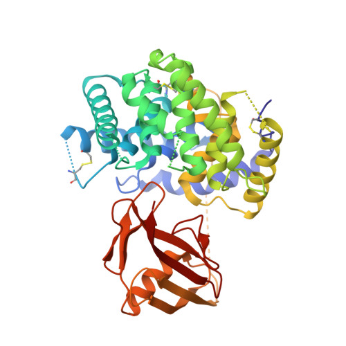
TC:CD320 in complex with nanobody TC-Nb4


Experimental Data Snapshot

  • Method: X-RAY DIFFRACTION
  • Resolution: 2.69 Å
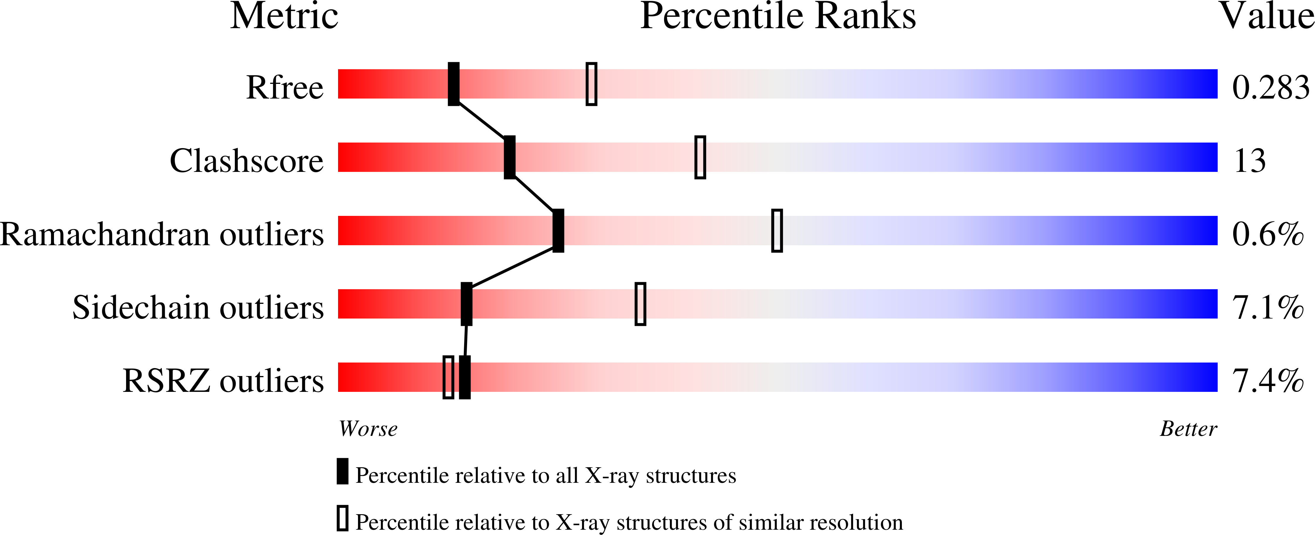
  • R-Value Free: 0.285 
  • R-Value Work: 0.227 
  • R-Value Observed: 0.230 

Starting Model: experimental
View more details

wwPDB Validation   3D Report Full Report


Ligand Structure Quality Assessment 


This is version 1.2 of the entry. See complete history


Literature

Generation of nanobodies targeting the human, transcobalamin-mediated vitamin B 12 uptake route.

Bloch, J.S.Sequeira, J.M.Ramirez, A.S.Quadros, E.V.Locher, K.P.

(2022) FASEB J 36: e22222-e22222

  • DOI: https://doi.org/10.1096/fj.202101376RR
  • Primary Citation of Related Structures:  
    7QBD, 7QBE, 7QBF, 7QBG

  • PubMed Abstract: 

    Cellular uptake of vitamin B 12 in humans is mediated by the endocytosis of the B 12 carrier protein transcobalamin (TC) via its cognate cell surface receptor TCblR, encoded by the CD320 gene. Because CD320 expression is associated with the cell cycle and upregulated in highly proliferating cells including cancer cells, this uptake route is a potential target for cancer therapy. We developed and characterized four camelid nanobodies that bind holo-TC (TC in complex with B 12 ) or the interface of the human holo-TC:TCblR complex with nanomolar affinities. We determined X-ray crystal structures of these nanobodies bound to holo-TC:TCblR, which enabled us to map their binding epitopes. When conjugated to the model toxin saporin, three of our nanobodies caused growth inhibition of HEK293T cells and therefore have the potential to inhibit the growth of human cancer cells. We visualized the cellular binding and endocytic uptake of the most potent nanobody (TC-Nb4) using fluorescent light microscopy. The co-crystal structure of holo-TC:TCblR with another nanobody (TC-Nb34) revealed novel features of the interface of TC and the LDLR-A1 domain of TCblR, rationalizing the decrease in the affinity of TC-B 12 binding caused by the Δ88 mutation in CD320.


  • Organizational Affiliation

    Institute of Molecular Biology and Biophysics, ETH Zürich, Zürich, Switzerland.


Macromolecules
Find similar proteins by:  (by identity cutoff)  |  3D Structure
Entity ID: 1
MoleculeChains Sequence LengthOrganismDetailsImage
Transcobalamin-2
A, C
409Homo sapiensMutation(s): 0 
Gene Names: TCN2TC2
UniProt & NIH Common Fund Data Resources
Find proteins for P20062 (Homo sapiens)
Explore P20062 
Go to UniProtKB:  P20062
PHAROS:  P20062
GTEx:  ENSG00000185339 
Entity Groups  
Sequence Clusters30% Identity50% Identity70% Identity90% Identity95% Identity100% Identity
UniProt GroupP20062
Sequence Annotations
Expand
  • Reference Sequence
Find similar proteins by:  (by identity cutoff)  |  3D Structure
Entity ID: 2
MoleculeChains Sequence LengthOrganismDetailsImage
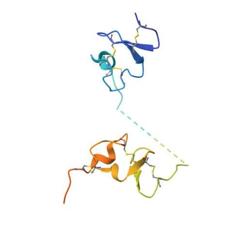
CD320 antigen
B, D
147Homo sapiensMutation(s): 0 
Gene Names: CD3208D6AUNQ198/PRO224
UniProt & NIH Common Fund Data Resources
Find proteins for Q9NPF0 (Homo sapiens)
Explore Q9NPF0 
Go to UniProtKB:  Q9NPF0
PHAROS:  Q9NPF0
GTEx:  ENSG00000167775 
Entity Groups  
Sequence Clusters30% Identity50% Identity70% Identity90% Identity95% Identity100% Identity
UniProt GroupQ9NPF0
Sequence Annotations
Expand
  • Reference Sequence
Find similar proteins by:  (by identity cutoff)  |  3D Structure
Entity ID: 3
MoleculeChains Sequence LengthOrganismDetailsImage
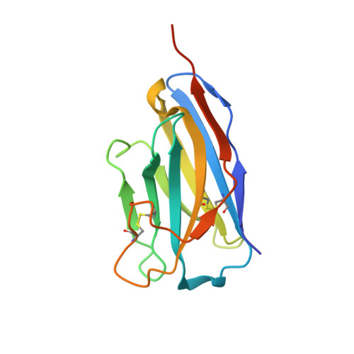
Anti-TC:CD320 nanobody TC-Nb4E,
F [auth G]
135Vicugna pacosMutation(s): 0 
Entity Groups  
Sequence Clusters30% Identity50% Identity70% Identity90% Identity95% Identity100% Identity
Sequence Annotations
Expand
  • Reference Sequence
Small Molecules
Ligands 3 Unique
IDChains Name / Formula / InChI Key2D Diagram3D Interactions
CNC
Query on CNC

Download Ideal Coordinates CCD File 
G [auth A],
L [auth C]
CYANOCOBALAMIN
C63 H89 Co N14 O14 P
SYZBZQWSWIJYAR-UVKKECPRSA-M
GOL
Query on GOL

Download Ideal Coordinates CCD File 
H [auth A],
I [auth A],
M [auth C]
GLYCEROL
C3 H8 O3
PEDCQBHIVMGVHV-UHFFFAOYSA-N
CA
Query on CA

Download Ideal Coordinates CCD File 
J [auth B]
K [auth B]
N [auth D]
O [auth D]
P [auth E]
J [auth B],
K [auth B],
N [auth D],
O [auth D],
P [auth E],
Q [auth G]
CALCIUM ION
Ca
BHPQYMZQTOCNFJ-UHFFFAOYSA-N
Experimental Data & Validation

Experimental Data

  • Method: X-RAY DIFFRACTION
  • Resolution: 2.69 Å
  • R-Value Free: 0.285 
  • R-Value Work: 0.227 
  • R-Value Observed: 0.230 
  • Space Group: P 21 21 21
Unit Cell:
Length ( Å )Angle ( ˚ )
a = 105.339α = 90
b = 116.418β = 90
c = 125.498γ = 90
Software Package:
Software NamePurpose
PHENIXrefinement
XDSdata reduction
XDSdata scaling
PHASERphasing

Structure Validation

View Full Validation Report


Ligand Structure Quality Assessment 


Entry History & Funding Information

Deposition Data


Funding OrganizationLocationGrant Number
Swiss National Science FoundationSwitzerland--

Revision History  (Full details and data files)

  • Version 1.0: 2022-03-16
    Type: Initial release
  • Version 1.1: 2024-01-31
    Changes: Data collection, Refinement description
  • Version 1.2: 2024-10-23
    Changes: Structure summary