7Q5U | pdb_00007q5u

The tandem SH2 domains of SYK with a bound CD3G diphospho-ITAM peptide


Experimental Data Snapshot

  • Method: X-RAY DIFFRACTION
  • Resolution: 2.40 Å
  • R-Value Free: 
    0.259 (Depositor), 0.260 (DCC) 
  • R-Value Work: 
    0.216 (Depositor), 0.220 (DCC) 

Starting Model: experimental
View more details

wwPDB Validation   3D Report Full Report


Ligand Structure Quality Assessment 

Created with Raphaël 2.3.0Worse 01 BetterLigand structure goodness of fit to experimental dataBest fitted P4GClick on this verticalbar to view details

This is version 1.4 of the entry. See complete history


Literature

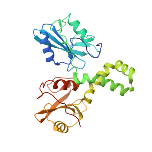
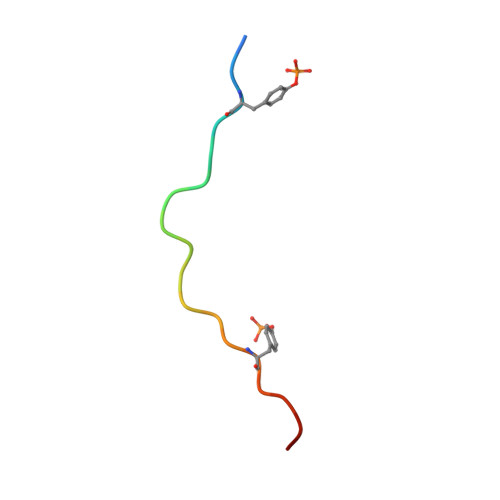
The mechanism of allosteric activation of SYK kinase derived from multiple phospho-ITAM-bound structures.

Bradshaw, W.J.Harris, G.Gileadi, O.Katis, V.L.

(2024) Structure 32: 2337-2351.e4

  • DOI: https://doi.org/10.1016/j.str.2024.09.024
  • Primary Citation of Related Structures:  
    7Q5T, 7Q5U, 7Q5W, 7Q63

  • PubMed Abstract: 

    Spleen tyrosine kinase (SYK) is central to adaptive and innate immune signaling. It features a regulatory region containing tandem SH2 (tSH2) domains separated by a helical "hinge" segment keeping SYK inactive by associating with the kinase domain. SYK activation is triggered when the tSH2 domains bind to a phosphorylated immunoreceptor tyrosine-based activation motif (ITAM) found on receptor tails. Past mutational studies have indicated that ITAM binding disrupts the hinge-kinase interaction, leading to SYK phosphorylation and activation. However, the mechanism of this process is unclear, as the ITAM interaction occurs far from the hinge region. We have determined crystal structures of three phospho-ITAMs in complex with the tSH2 domains, revealing a highly conserved binding mechanism. These structures, together with mutational studies and biophysical analyses, reveal that phospho-ITAM binding restricts SH2 domain movement and causes allosteric changes in the hinge region. These changes are not compatible with the association of the kinase domain, leading to kinase activation.


  • Organizational Affiliation

    Alzheimer's Research UK Oxford Drug Discovery Institute, Centre for Medicines Discovery, Nuffield Department of Medicine Research Building, Old Road Campus, University of Oxford, Oxford OX3 7FZ, UK.


Macromolecules
Find similar proteins by:  (by identity cutoff)  |  3D Structure
Entity ID: 1
MoleculeChains Sequence LengthOrganismDetailsImage
Tyrosine-protein kinase SYK265Homo sapiensMutation(s): 0 
Gene Names: SYK
EC: 2.7.10.2
UniProt & NIH Common Fund Data Resources
Find proteins for P43405 (Homo sapiens)
Explore P43405 
Go to UniProtKB:  P43405
PHAROS:  P43405
GTEx:  ENSG00000165025 
Entity Groups  
Sequence Clusters30% Identity50% Identity70% Identity90% Identity95% Identity100% Identity
UniProt GroupP43405
Sequence Annotations
Expand
  • Reference Sequence

Find similar proteins by:  Sequence   |   3D Structure  

Entity ID: 2
MoleculeChains Sequence LengthOrganismDetailsImage
T-cell surface glycoprotein CD3 gamma chain20Homo sapiensMutation(s): 0 
UniProt & NIH Common Fund Data Resources
Find proteins for P09693 (Homo sapiens)
Explore P09693 
Go to UniProtKB:  P09693
PHAROS:  P09693
GTEx:  ENSG00000160654 
Entity Groups  
Sequence Clusters30% Identity50% Identity70% Identity90% Identity95% Identity100% Identity
UniProt GroupP09693
Sequence Annotations
Expand
  • Reference Sequence
Small Molecules
Ligands 3 Unique
IDChains Name / Formula / InChI Key2D Diagram3D Interactions
P4G
Query on P4G

Download Ideal Coordinates CCD File 
P [auth AAA],
V [auth EEE]
1-ETHOXY-2-(2-ETHOXYETHOXY)ETHANE
C8 H18 O3
RRQYJINTUHWNHW-UHFFFAOYSA-N
PEG
Query on PEG

Download Ideal Coordinates CCD File 
O [auth AAA],
Q [auth BBB],
U [auth EEE],
W [auth FFF]
DI(HYDROXYETHYL)ETHER
C4 H10 O3
MTHSVFCYNBDYFN-UHFFFAOYSA-N
EDO
Query on EDO

Download Ideal Coordinates CCD File 
M [auth AAA],
N [auth AAA],
R [auth CCC],
S [auth CCC],
T [auth DDD]
1,2-ETHANEDIOL
C2 H6 O2
LYCAIKOWRPUZTN-UHFFFAOYSA-N
Modified Residues  1 Unique
IDChains TypeFormula2D DiagramParent
PTR
Query on PTR
G [auth GGG]
H [auth HHH]
I [auth III]
J [auth JJJ]
K [auth KKK]
L-PEPTIDE LINKINGC9 H12 N O6 PTYR
Experimental Data & Validation

Experimental Data

  • Method: X-RAY DIFFRACTION
  • Resolution: 2.40 Å
  • R-Value Free:  0.259 (Depositor), 0.260 (DCC) 
  • R-Value Work:  0.216 (Depositor), 0.220 (DCC) 
Space Group: P 1 21 1
Unit Cell:
Length ( Å )Angle ( ˚ )
a = 92.321α = 90
b = 129.312β = 100.232
c = 95.346γ = 90
Software Package:
Software NamePurpose
REFMACrefinement
DIALSdata reduction
Aimlessdata scaling
PHASERphasing

Structure Validation

View Full Validation Report


Ligand Structure Quality Assessment 

Created with Raphaël 2.3.0Worse 01 BetterLigand structure goodness of fit to experimental dataBest fitted P4GClick on this verticalbar to view details

Entry History & Funding Information

Deposition Data


Funding OrganizationLocationGrant Number
National Institutes of Health/National Institute on Aging (NIH/NIA)United States5U54AG065187-03

Revision History  (Full details and data files)

  • Version 1.0: 2021-11-24
    Type: Initial release
  • Version 1.1: 2024-01-31
    Changes: Data collection, Derived calculations, Refinement description
  • Version 1.2: 2024-10-23
    Changes: Structure summary
  • Version 1.3: 2024-10-30
    Changes: Database references
  • Version 1.4: 2025-01-29
    Changes: Database references