7Q5N | pdb_00007q5n

Crystal structure of Chaetomium thermophilum Ahp1-Urm1 complex


Experimental Data Snapshot

  • Method: X-RAY DIFFRACTION
  • Resolution: 2.50 Å
  • R-Value Free: 
    0.272 (Depositor), 0.280 (DCC) 
  • R-Value Work: 
    0.234 (Depositor), 0.244 (DCC) 
  • R-Value Observed: 
    0.236 (Depositor) 

Starting Model: experimental
View more details

wwPDB Validation 3D Report Full Report

Validation slider image for 7Q5N

This is version 1.5 of the entry. See complete history

Literature

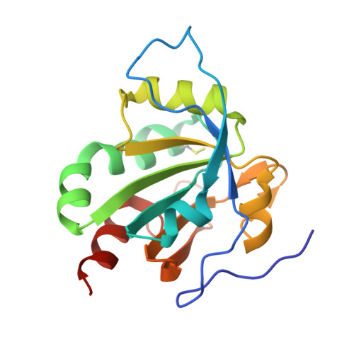
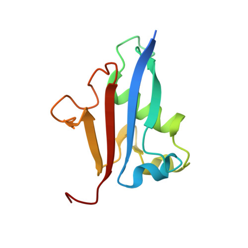
E2/E3-independent ubiquitin-like protein conjugation by Urm1 is directly coupled to cysteine persulfidation.

Ravichandran, K.E.Kaduhr, L.Skupien-Rabian, B.Shvetsova, E.Sokolowski, M.Krutyholowa, R.C.Kwasna, D.Brachmann, C.Lin, S.Guzman Perez, S.Wilk, P.Kosters, M.Grudnik, P.Jankowska, U.Leidel, S.A.Schaffrath, R.Glatt, S.

(2022) EMBO J 41: e111318-e111318

  • DOI: https://doi.org/10.15252/embj.2022111318
  • Primary Citation Related Structures: 
    7Q5N, 7Q68, 7Q69, 7Q6A

  • PubMed Abstract: 

    Post-translational modifications by ubiquitin-like proteins (UBLs) are essential for nearly all cellular processes. Ubiquitin-related modifier 1 (Urm1) is a unique UBL, which plays a key role in tRNA anticodon thiolation as a sulfur carrier protein (SCP) and is linked to the noncanonical E1 enzyme Uba4 (ubiquitin-like protein activator 4). While Urm1 has also been observed to conjugate to target proteins like other UBLs, the molecular mechanism of its attachment remains unknown. Here, we reconstitute the covalent attachment of thiocarboxylated Urm1 to various cellular target proteins in vitro, revealing that, unlike other known UBLs, this process is E2/E3-independent and requires oxidative stress. Furthermore, we present the crystal structures of the peroxiredoxin Ahp1 before and after the covalent attachment of Urm1. Surprisingly, we show that urmylation is accompanied by the transfer of sulfur to cysteine residues in the target proteins, also known as cysteine persulfidation. Our results illustrate the role of the Uba4-Urm1 system as a key evolutionary link between prokaryotic SCPs and the UBL modifications observed in modern eukaryotes.


  • Organizational Affiliation
    • Malopolska Centre of Biotechnology (MCB), Jagiellonian University, Krakow, Poland.

Macromolecule Content 

  • Total Structure Weight: 186.54 kDa 
  • Atom Count: 12,664 
  • Modeled Residue Count: 1,641 
  • Deposited Residue Count: 1,710 
  • Unique protein chains: 2

Macromolecules

Find similar proteins by:|  3D Structure
Entity ID: 1
MoleculeChains  Sequence LengthOrganismDetailsImage
Thioredoxin domain-containing protein
A, B, C, D, E
A, B, C, D, E, F
174Thermochaetoides thermophila DSM 1495Mutation(s): 1 
Gene Names: CTHT_0014370
EC: 1.11.1.24
UniProt
Find proteins for G0S1P8 (Chaetomium thermophilum (strain DSM 1495 / CBS 144.50 / IMI 039719))
Explore G0S1P8 
Go to UniProtKB:  G0S1P8
Entity Groups
Sequence Clusters30% Identity50% Identity70% Identity90% Identity95% Identity100% Identity
UniProt GroupG0S1P8
Sequence Annotations
Expand
Reference Sequence
Find similar proteins by:|  3D Structure
Entity ID: 2
MoleculeChains  Sequence LengthOrganismDetailsImage
Ubiquitin-related modifier 1
G, H, I, J, K
G, H, I, J, K, L
111Thermochaetoides thermophila DSM 1495Mutation(s): 1 
Gene Names: URM1CTHT_0062030
UniProt
Find proteins for G0SE11 (Chaetomium thermophilum (strain DSM 1495 / CBS 144.50 / IMI 039719))
Explore G0SE11 
Go to UniProtKB:  G0SE11
Entity Groups
Sequence Clusters30% Identity50% Identity70% Identity90% Identity95% Identity100% Identity
UniProt GroupG0SE11
Sequence Annotations
Expand
Reference Sequence

Small Molecules

Ligands 1 Unique
IDChains Name / Formula / InChI Key2D Diagram3D Interactions
ZN

Query on ZN


Download:Ideal Coordinates CCD File
AA [auth G]
AB [auth L]
BA [auth H]
CA [auth H]
DA [auth H]
AA [auth G],
AB [auth L],
BA [auth H],
CA [auth H],
DA [auth H],
EA [auth H],
FA [auth H],
GA [auth H],
HA [auth H],
IA [auth H],
JA [auth I],
KA [auth I],
LA [auth I],
M [auth A],
MA [auth I],
N [auth B],
NA [auth I],
O [auth B],
OA [auth J],
P [auth B],
PA [auth J],
Q [auth C],
QA [auth J],
R [auth C],
RA [auth J],
S [auth C],
SA [auth K],
T [auth D],
TA [auth K],
U [auth D],
UA [auth K],
V [auth E],
VA [auth K],
W [auth E],
WA [auth K],
X [auth E],
XA [auth K],
Y [auth F],
YA [auth L],
Z [auth F],
ZA [auth L]
ZINC ION
Zn
PTFCDOFLOPIGGS-UHFFFAOYSA-N
Modified Residues  1 Unique
IDChains TypeFormula2D DiagramParent
CSS
Query on CSS
A, B, C, D, E
A, B, C, D, E, F
L-PEPTIDE LINKINGC3 H7 N O2 S2CYS

Experimental Data & Validation

Experimental Data

  • Method: X-RAY DIFFRACTION
  • Resolution: 2.50 Å
  • R-Value Free:  0.272 (Depositor), 0.280 (DCC) 
  • R-Value Work:  0.234 (Depositor), 0.244 (DCC) 
  • R-Value Observed: 0.236 (Depositor) 
Space Group: C 2 2 21
Unit Cell:
Length ( Å )Angle ( ˚ )
a = 185.43α = 90
b = 197.22β = 90
c = 139.18γ = 90
Software Package:
Software NamePurpose
REFMACrefinement
PHENIXrefinement
XDSdata reduction
XSCALEdata scaling
PHASERphasing

Structure Validation

View Full Validation Report


Entry History 

& Funding Information

Deposition Data


Funding OrganizationLocationGrant Number
Foundation for Polish SciencePolandTEAM TECH CORE FACILITY/2017-4/6
European Research Council (ERC)Poland101001394
Polish National Science CentrePoland2018/31/B/NZ1/03559
Foundation for Polish SciencePolandFirstTEAM/2016-1/2

Revision History  (Full details and data files)

  • Version 1.0: 2022-08-24
    Type: Initial release
  • Version 1.1: 2022-08-31
    Changes: Database references
  • Version 1.2: 2022-09-28
    Changes: Database references
  • Version 1.3: 2022-10-26
    Changes: Database references
  • Version 1.4: 2024-01-31
    Changes: Data collection, Refinement description
  • Version 1.5: 2024-10-23
    Changes: Structure summary