7Q0X | pdb_00007q0x

Bovine Trypsin co-crystallized with V(IV)OSO4 and pic


Experimental Data Snapshot

  • Method: X-RAY DIFFRACTION
  • Resolution: 1.09 Å
  • R-Value Free: 
    0.136 (Depositor), 0.161 (DCC) 
  • R-Value Work: 
    0.111 (Depositor) 
  • R-Value Observed: 
    0.113 (Depositor) 

Starting Model: experimental
View more details

wwPDB Validation 3D Report Full Report

Validation slider image for 7Q0X

Ligand Structure Quality Assessment 


This is version 1.5 of the entry. See complete history

Literature

Binding of V IV O 2+ , V IV OL, V IV OL 2 and V V O 2 L Moieties to Proteins: X-ray/Theoretical Characterization and Biological Implications.

Santos, M.F.A.Sciortino, G.Correia, I.Fernandes, A.C.P.Santos-Silva, T.Pisanu, F.Garribba, E.Costa Pessoa, J.

(2022) Chemistry 28: e202200105-e202200105

  • DOI: https://doi.org/10.1002/chem.202200105
  • Primary Citation Related Structures: 
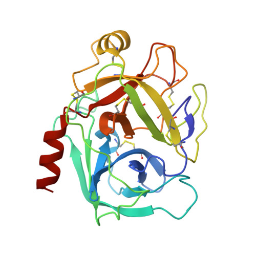
    7Q0T, 7Q0U, 7Q0V, 7Q0W, 7Q0X

  • PubMed Abstract: 

    Vanadium compounds have frequently been proposed as therapeutics, but their application has been hampered by the lack of information on the different V-containing species that may form and how these interact with blood and cell proteins, and with enzymes. Herein, we report several resolved crystal structures of lysozyme with bound V IV O 2+ and V IV OL 2+ , where L=2,2'-bipyridine or 1,10-phenanthroline (phen), and of trypsin with V IV O(picolinato) 2 and V V O 2 (phen) + moieties. Computational studies complete the refinement and shed light on the relevant role of hydrophobic interactions, hydrogen bonds, and microsolvation in stabilizating the structure. Noteworthy is that the trypsin-V V O 2 (phen) and trypsin-V IV O(OH)(phen) adducts correspond to similar energies, thus suggesting a possible interconversion under physiological/biological conditions. The obtained data support the relevance of hydrolysis of V IV and V V complexes in the several types of binding established with proteins and the formation of different adducts that might contribute to their pharmacological action, and significantly widen our knowledge of vanadium-protein interactions.


  • Organizational Affiliation
    • Centro de Química Estrutural and Departamento de Engenharia Química, Institute of Molecular Sciences, Instituto Superior Técnico, Universidade de Lisboa, Av. Rovisco Pais, 1049-001, Lisboa, Portugal.

Macromolecule Content 

  • Total Structure Weight: 24.23 kDa 
  • Atom Count: 2,002 
  • Modeled Residue Count: 223 
  • Deposited Residue Count: 223 
  • Unique protein chains: 1

Macromolecules

Find similar proteins by:|  3D Structure
Entity ID: 1
MoleculeChains  Sequence LengthOrganismDetailsImage
Cationic trypsin223Bos taurusMutation(s): 0 
EC: 3.4.21.4
UniProt
Find proteins for P00760 (Bos taurus)
Explore P00760 
Go to UniProtKB:  P00760
Entity Groups
Sequence Clusters30% Identity50% Identity70% Identity90% Identity95% Identity100% Identity
UniProt GroupP00760
Sequence Annotations
Expand
Reference Sequence

Small Molecules

Ligands 6 Unique
IDChains Name / Formula / InChI Key2D Diagram3D Interactions
6PC
(Subject of Investigation/LOI)

Query on 6PC


Download:Ideal Coordinates CCD File
D [auth A],
E [auth A]
PYRIDINE-2-CARBOXYLIC ACID
C6 H5 N O2
SIOXPEMLGUPBBT-UHFFFAOYSA-N
SO4

Query on SO4


Download:Ideal Coordinates CCD File
K [auth A]SULFATE ION
O4 S
QAOWNCQODCNURD-UHFFFAOYSA-L
GOL

Query on GOL


Download:Ideal Coordinates CCD File
F [auth A],
G [auth A],
H [auth A],
I [auth A],
J [auth A]
GLYCEROL
C3 H8 O3
PEDCQBHIVMGVHV-UHFFFAOYSA-N
V
(Subject of Investigation/LOI)

Query on V


Download:Ideal Coordinates CCD File
B [auth A]VANADIUM ION
V
KOKKJWHERHSKEB-UHFFFAOYSA-N
CA

Query on CA


Download:Ideal Coordinates CCD File
L [auth A]CALCIUM ION
Ca
BHPQYMZQTOCNFJ-UHFFFAOYSA-N
O

Query on O


Download:Ideal Coordinates CCD File
C [auth A]OXYGEN ATOM
O
XLYOFNOQVPJJNP-UHFFFAOYSA-N

Experimental Data & Validation

Experimental Data

  • Method: X-RAY DIFFRACTION
  • Resolution: 1.09 Å
  • R-Value Free:  0.136 (Depositor), 0.161 (DCC) 
  • R-Value Work:  0.111 (Depositor) 
  • R-Value Observed: 0.113 (Depositor) 
Space Group: P 31 2 1
Unit Cell:
Length ( Å )Angle ( ˚ )
a = 54.522α = 90
b = 54.522β = 90
c = 108.199γ = 120
Software Package:
Software NamePurpose
REFMACrefinement
XDSdata reduction
Aimlessdata scaling
PHASERphasing

Structure Validation

View Full Validation Report


Ligand Structure Quality Assessment 


Entry History 

& Funding Information

Deposition Data


Funding OrganizationLocationGrant Number
Fundacao para a Ciencia e a TecnologiaPortugal--

Revision History  (Full details and data files)

  • Version 1.0: 2022-05-25
    Type: Initial release
  • Version 1.1: 2022-06-01
    Changes: Database references
  • Version 1.2: 2022-06-15
    Changes: Database references
  • Version 1.3: 2022-07-27
    Changes: Database references
  • Version 1.4: 2024-01-31
    Changes: Data collection, Refinement description
  • Version 1.5: 2024-10-23
    Changes: Structure summary