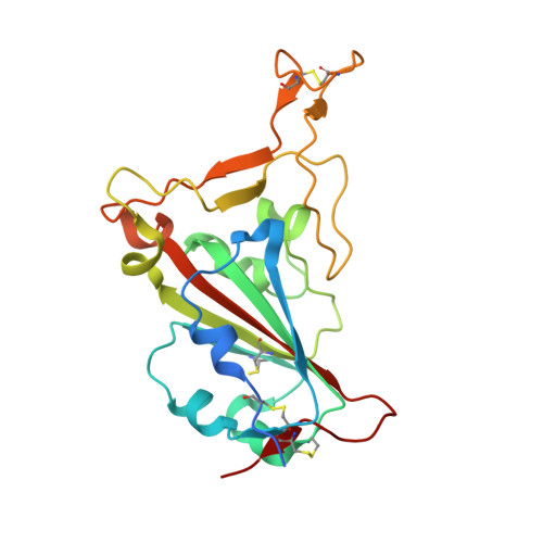
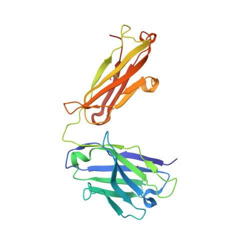
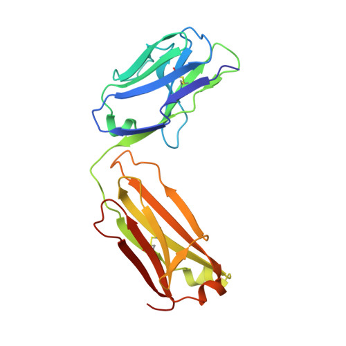
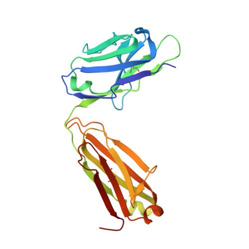
Alpha-B.1.1.7, Beta-B.1.351, Gamma-P.1, and Delta-B.1.617.2 variants of SARS-CoV-2 express multiple mutations in the spike protein (S). These may alter the antigenic structure of S, causing escape from natural or vaccine-induced immunity. Beta is particularly difficult to neutralize using serum induced by early pandemic SARS-CoV-2 strains and is most antigenically separated from Delta. To understand this, we generated 674 mAbs from Beta-infected individuals and performed a detailed structure-function analysis of the 27 most potent mAbs: one binding the spike N-terminal domain (NTD), the rest the receptor-binding domain (RBD). Two of these RBD-binding mAbs recognize a neutralizing epitope conserved between SARS-CoV-1 and -2, while 18 target mutated residues in Beta: K417N, E484K, and N501Y. There is a major response to N501Y, including a public IgVH4-39 sequence, with E484K and K417N also targeted. Recognition of these key residues underscores why serum from Beta cases poorly neutralizes early pandemic and Delta viruses.
Organizational Affiliation:
Wellcome Centre for Human Genetics, Nuffield Department of Medicine, University of Oxford, Oxford, UK; Chinese Academy of Medical Science Oxford Institute, University of Oxford, Oxford, UK.
Chinese Academy of Medical Science Oxford Institute, University of Oxford, Oxford, UK; Division of Structural Biology, Nuffield Department of Medicine, University of Oxford, The Wellcome Centre for Human Genetics, Oxford, UK.
Wellcome Centre for Human Genetics, Nuffield Department of Medicine, University of Oxford, Oxford, UK.
Division of Structural Biology, Nuffield Department of Medicine, University of Oxford, The Wellcome Centre for Human Genetics, Oxford, UK.
Wellcome Centre for Human Genetics, Nuffield Department of Medicine, University of Oxford, Oxford, UK; Oxford University Hospitals NHS Foundation Trust, Oxford, UK.
Oxford University Hospitals NHS Foundation Trust, Oxford, UK; Peter Medawar Building for Pathogen Research, Nuffield Department of Medicine, University of Oxford, Oxford, UK; Nuffield Department of Clinical Neurosciences, University of Oxford, Oxford, UK.
Department of Medicine, Washington University School of Medicine, St. Louis, MO 63110, USA; Department of Pathology and Immunology, Washington University School of Medicine, St. Louis, MO 63110, USA.
Oxford University Hospitals NHS Foundation Trust, Oxford, UK; Peter Medawar Building for Pathogen Research, Nuffield Department of Medicine, University of Oxford, Oxford, UK.
Oxford University Hospitals NHS Foundation Trust, Oxford, UK.
National Infection Service, Public Health England (PHE), Porton Down, Salisbury, UK.
Viral Pseudotype Unit, Medway School of Pharmacy, University of Kent and Greenwich, Chatham Maritime, Kent ME4 4TB, UK.
Peter Medawar Building for Pathogen Research, Nuffield Department of Medicine, University of Oxford, Oxford, UK; Department of Paediatrics, University of Oxford, Oxford, UK.
Department of Medicine, Washington University School of Medicine, St. Louis, MO 63110, USA; Department of Pathology and Immunology, Washington University School of Medicine, St. Louis, MO 63110, USA; Department of Molecular Microbiology, Washington University School of Medicine, St. Louis, MO 63110, USA; The Andrew M. and Jane M. Bursky Center for Human Immunology and Immunotherapy Programs, Washington University School of Medicine, St. Louis, MO 63110, USA.
Wellcome Centre for Human Genetics, Nuffield Department of Medicine, University of Oxford, Oxford, UK; Chinese Academy of Medical Science Oxford Institute, University of Oxford, Oxford, UK; Siriraj Center of Research Excellence in Dengue & Emerging Pathogens, Dean Office for Research, Faculty of Medicine Siriraj Hospital, Mahidol University, Bangkok, Thailand. Electronic address: jmongkol@well.ox.ac.uk.
Division of Structural Biology, Nuffield Department of Medicine, University of Oxford, The Wellcome Centre for Human Genetics, Oxford, UK. Electronic address: ren@strubi.ox.ac.uk.
Wellcome Centre for Human Genetics, Nuffield Department of Medicine, University of Oxford, Oxford, UK; Chinese Academy of Medical Science Oxford Institute, University of Oxford, Oxford, UK; Division of Structural Biology, Nuffield Department of Medicine, University of Oxford, The Wellcome Centre for Human Genetics, Oxford, UK; Diamond Light Source Ltd, Harwell Science & Innovation Campus, Didcot, UK; Instruct-ERIC, Oxford House, Parkway Court, John Smith Drive, Oxford, UK. Electronic address: dave@strubi.ox.ac.uk.
Wellcome Centre for Human Genetics, Nuffield Department of Medicine, University of Oxford, Oxford, UK; Chinese Academy of Medical Science Oxford Institute, University of Oxford, Oxford, UK. Electronic address: gavin.screaton@medsci.ox.ac.uk.
AA [auth L] G [auth E] H [auth E] I [auth E] IA [auth B]
AA [auth L], G [auth E], H [auth E], I [auth E], IA [auth B], J [auth E], JA [auth B], KA [auth B], P [auth H], Q [auth H], R [auth H], SA [auth A], T [auth H]
DA [auth L] EA [auth L] FA [auth L] GA [auth L] HA [auth L]
DA [auth L], EA [auth L], FA [auth L], GA [auth L], HA [auth L], O [auth E], OA [auth B], PA [auth B], V [auth H], W [auth H], X [auth H], XA [auth A], Y [auth H], Z [auth H]
BA [auth L] CA [auth L] K [auth E] L [auth E] LA [auth B]
BA [auth L], CA [auth L], K [auth E], L [auth E], LA [auth B], M [auth E], MA [auth B], N [auth E], NA [auth B], TA [auth A], U [auth H], UA [auth A], VA [auth A], WA [auth A]