7Q07 | pdb_00007q07

Ketol-acid reductoisomerase from Methanothermococcus thermolithotrophicus in the open state with NADP and tartrate


Experimental Data Snapshot

  • Method: X-RAY DIFFRACTION
  • Resolution: 2.20 Å
  • R-Value Free: 
    0.212 (Depositor), 0.229 (DCC) 
  • R-Value Work: 
    0.186 (Depositor), 0.206 (DCC) 
  • R-Value Observed: 
    0.190 (Depositor) 

Starting Model: experimental
View more details

wwPDB Validation 3D Report Full Report

Validation slider image for 7Q07

Ligand Structure Quality Assessment 


This is version 1.1 of the entry. See complete history

Literature

Structural Rearrangements of a Dodecameric Ketol-Acid Reductoisomerase Isolated from a Marine Thermophilic Methanogen.

Lemaire, O.N.Muller, M.C.Kahnt, J.Wagner, T.

(2021) Biomolecules 11

  • DOI: https://doi.org/10.3390/biom11111679
  • Primary Citation Related Structures: 
    7Q03, 7Q07

  • PubMed Abstract: 

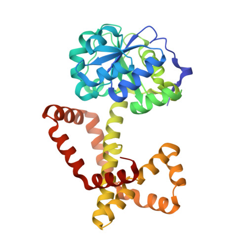
    Ketol-acid reductoisomerase (KARI) orchestrates the biosynthesis of branched-chain amino acids, an elementary reaction in prototrophic organisms as well as a valuable process in biotechnology. Bacterial KARIs belonging to class I organise as dimers or dodecamers and were intensively studied to understand their remarkable specificity towards NADH or NADPH, but also to develop antibiotics. Here, we present the first structural study on a KARI natively isolated from a methanogenic archaea. The dodecameric structure of 0.44-MDa was obtained in two different conformations, an open and close state refined to a resolution of 2.2-Å and 2.1-Å, respectively. These structures illustrate the conformational movement required for substrate and coenzyme binding. While the close state presents the complete NADP bound in front of a partially occupied Mg 2+ -site, the Mg 2+ -free open state contains a tartrate at the nicotinamide location and a bound NADP with the adenine-nicotinamide protruding out of the active site. Structural comparisons show a very high conservation of the active site environment and detailed analyses point towards few specific residues required for the dodecamerisation. These residues are not conserved in other dodecameric KARIs that stabilise their trimeric interface differently, suggesting that dodecamerisation, the cellular role of which is still unknown, might have occurred several times in the evolution of KARIs.


  • Organizational Affiliation
    • Microbial Metabolism Research Group, Max Planck Institute for Marine Microbiology, Celsiusstraße 1, 28359 Bremen, Germany.

Macromolecule Content 

  • Total Structure Weight: 37.62 kDa 
  • Atom Count: 2,725 
  • Modeled Residue Count: 327 
  • Deposited Residue Count: 330 
  • Unique protein chains: 1

Macromolecules

Find similar proteins by:|  3D Structure
Entity ID: 1
MoleculeChains  Sequence LengthOrganismDetailsImage
Ketol-Acid Reductoisomerase from Methanothermococcus thermolithotrophicus330Methanothermococcus thermolithotrophicus DSM 2095Mutation(s): 0 
EC: 1.1.1.86
UniProt
Find proteins for A0A8X6EH16 (Methanothermococcus thermolithotrophicus DSM 2095)
Explore A0A8X6EH16 
Go to UniProtKB:  A0A8X6EH16
Entity Groups
Sequence Clusters30% Identity50% Identity70% Identity90% Identity95% Identity100% Identity
UniProt GroupA0A8X6EH16
Sequence Annotations
Expand
Reference Sequence

Small Molecules

Ligands 4 Unique
IDChains Name / Formula / InChI Key2D Diagram3D Interactions
NAP
(Subject of Investigation/LOI)

Query on NAP


Download:Ideal Coordinates CCD File
E [auth A]NADP NICOTINAMIDE-ADENINE-DINUCLEOTIDE PHOSPHATE
C21 H28 N7 O17 P3
XJLXINKUBYWONI-NNYOXOHSSA-N
TLA

Query on TLA


Download:Ideal Coordinates CCD File
B [auth A]L(+)-TARTARIC ACID
C4 H6 O6
FEWJPZIEWOKRBE-JCYAYHJZSA-N
EDO

Query on EDO


Download:Ideal Coordinates CCD File
D [auth A]1,2-ETHANEDIOL
C2 H6 O2
LYCAIKOWRPUZTN-UHFFFAOYSA-N
CL

Query on CL


Download:Ideal Coordinates CCD File
C [auth A],
F [auth A],
G [auth A]
CHLORIDE ION
Cl
VEXZGXHMUGYJMC-UHFFFAOYSA-M

Experimental Data & Validation

Experimental Data

  • Method: X-RAY DIFFRACTION
  • Resolution: 2.20 Å
  • R-Value Free:  0.212 (Depositor), 0.229 (DCC) 
  • R-Value Work:  0.186 (Depositor), 0.206 (DCC) 
  • R-Value Observed: 0.190 (Depositor) 
Space Group: I 2 3
Unit Cell:
Length ( Å )Angle ( ˚ )
a = 130.827α = 90
b = 130.827β = 90
c = 130.827γ = 90
Software Package:
Software NamePurpose
PHENIXrefinement
PDB_EXTRACTdata extraction
XDSdata reduction
SCALAdata scaling
PHENIXphasing

Structure Validation

View Full Validation Report


Ligand Structure Quality Assessment 


Entry History 

& Funding Information

Deposition Data


Funding OrganizationLocationGrant Number
Max Planck SocietyGermany--

Revision History  (Full details and data files)

  • Version 1.0: 2021-12-08
    Type: Initial release
  • Version 1.1: 2024-01-31
    Changes: Data collection, Derived calculations, Refinement description