7PRS

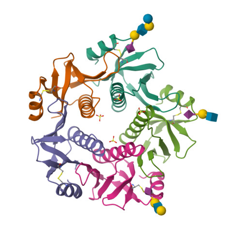
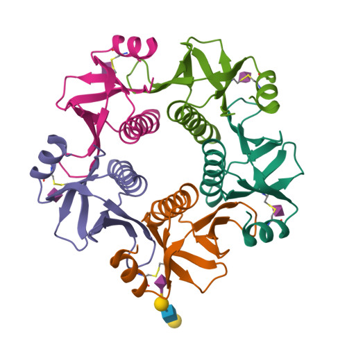
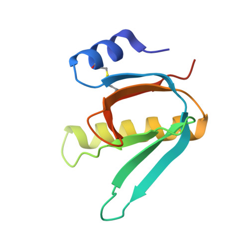
Crystal Structure of the B subunit of heat labile enterotoxin LT-IIc from Escherichia coli in complex with Sialyl-lacto-N-neotetraose d


Experimental Data Snapshot

  • Method: X-RAY DIFFRACTION
  • Resolution: 2.00 Å
  • R-Value Free: 0.230 
  • R-Value Work: 0.188 

Starting Model: experimental
View more details

wwPDB Validation   3D Report Full Report


Ligand Structure Quality Assessment 


This is version 1.3 of the entry. See complete history


Literature

Characterization of the ganglioside recognition profile of Escherichia coli heat-labile enterotoxin LT-IIc.

Zalem, D.Juhas, M.Terrinoni, M.King-Lyons, N.Lebens, M.Varrot, A.Connell, T.D.Teneberg, S.

(2022) Glycobiology 32: 391-403

  • DOI: https://doi.org/10.1093/glycob/cwab133
  • Primary Citation of Related Structures:  
    7PRP, 7PRS

  • PubMed Abstract: 

    The heat-labile enterotoxins of Escherichia coli and cholera toxin of Vibrio cholerae are related in structure and function. Each of these oligomeric toxins is comprised of one A polypeptide and five B polypeptides. The B-subunits bind to gangliosides, which are followed by uptake into the intoxicated cell and activation of the host's adenylate cyclase by the A-subunits. There are two antigenically distinct groups of these toxins. Group I includes cholera toxin and type I heat-labile enterotoxin of E. coli; group II contains the type II heat-labile enterotoxins of E. coli. Three variants of type II toxins, designated LT-IIa, LT-IIb and LT-IIc have been described. Earlier studies revealed the crystalline structure of LT-IIb. Herein the carbohydrate binding specificity of LT-IIc B-subunits was investigated by glycosphingolipid binding studies on thin-layer chromatograms and in microtiter wells. Binding studies using a large variety of glycosphingolipids showed that LT-IIc binds with high affinity to gangliosides with a terminal Neu5Acα3Gal or Neu5Gcα3Gal, e.g. the gangliosides GM3, GD1a and Neu5Acα3-/Neu5Gcα3--neolactotetraosylceramide and Neu5Acα3-/Neu5Gcα3-neolactohexaosylceramide. The crystal structure of LT-IIc B-subunits alone and with bound LSTd/sialyl-lacto-N-neotetraose d pentasaccharide uncovered the molecular basis of the ganglioside recognition. These studies revealed common and unique functional structures of the type II family of heat-labile enterotoxins.


  • Organizational Affiliation

    Department of Medical Biochemistry and Cell Biology, Sahlgrenska Academy, Institute of Biomedicine, University of Gothenburg, Sweden.


Macromolecules
Find similar proteins by:  (by identity cutoff)  |  3D Structure
Entity ID: 1
MoleculeChains Sequence LengthOrganismDetailsImage
Heat-labile enterotoxin IIA, B chain104Escherichia coliMutation(s): 0 
Gene Names: LT-IIc1 BGQM13_12885HVV37_03950HVV78_10285HVW37_13205
UniProt
Find proteins for H6W8F2 (Escherichia coli)
Explore H6W8F2 
Go to UniProtKB:  H6W8F2
Entity Groups  
Sequence Clusters30% Identity50% Identity70% Identity90% Identity95% Identity100% Identity
UniProt GroupH6W8F2
Sequence Annotations
Expand
  • Reference Sequence
Oligosaccharides

Help

Entity ID: 2
MoleculeChains Length2D Diagram Glycosylation3D Interactions
N-acetyl-alpha-neuraminic acid-(2-3)-beta-D-galactopyranose-(1-4)-2-acetamido-2-deoxy-beta-D-glucopyranose-(1-3)-beta-D-galactopyranose-(1-4)-alpha-D-glucopyranoseK [auth DaD]5N/A
Entity ID: 3
MoleculeChains Length2D Diagram Glycosylation3D Interactions
N-acetyl-alpha-neuraminic acid-(2-3)-beta-D-galactopyranose-(1-4)-2-acetamido-2-deoxy-beta-D-glucopyranose-(1-3)-beta-D-galactopyranoseL [auth GaG],
N [auth JaJ]
4N/A
Glycosylation Resources
GlyTouCan:  G69440RD
GlyCosmos:  G69440RD
GlyGen:  G69440RD
Entity ID: 4
MoleculeChains Length2D Diagram Glycosylation3D Interactions
N-acetyl-alpha-neuraminic acid-(2-3)-beta-D-galactopyranose-(1-4)-2-acetamido-2-deoxy-beta-D-glucopyranoseM [auth HaH]3N/A
Glycosylation Resources
GlyTouCan:  G00065MO
GlyCosmos:  G00065MO
GlyGen:  G00065MO
Small Molecules
Ligands 2 Unique
IDChains Name / Formula / InChI Key2D Diagram3D Interactions
SIA (Subject of Investigation/LOI)
Query on SIA

Download Ideal Coordinates CCD File 
S [auth III],
T [auth JJJ],
U [auth KKK],
V [auth LLL]
N-acetyl-alpha-neuraminic acid
C11 H19 N O9
SQVRNKJHWKZAKO-YRMXFSIDSA-N
SO4
Query on SO4

Download Ideal Coordinates CCD File 
O [auth DDD],
P [auth EEE],
Q [auth FFF],
R [auth HHH]
SULFATE ION
O4 S
QAOWNCQODCNURD-UHFFFAOYSA-L
Experimental Data & Validation

Experimental Data

  • Method: X-RAY DIFFRACTION
  • Resolution: 2.00 Å
  • R-Value Free: 0.230 
  • R-Value Work: 0.188 
  • Space Group: P 21 21 21
Unit Cell:
Length ( Å )Angle ( ˚ )
a = 73.09α = 90
b = 121.269β = 90
c = 126.732γ = 90
Software Package:
Software NamePurpose
REFMACrefinement
XDSdata reduction
Aimlessdata scaling
PHASERphasing

Structure Validation

View Full Validation Report


Ligand Structure Quality Assessment 


Entry History & Funding Information

Deposition Data

  • Released Date: 2022-02-02 
  • Deposition Author(s): Varrot, A.

Funding OrganizationLocationGrant Number
European CommissionEuropean Union653706

Revision History  (Full details and data files)

  • Version 1.0: 2022-02-02
    Type: Initial release
  • Version 1.1: 2022-05-04
    Changes: Database references
  • Version 1.2: 2024-01-31
    Changes: Data collection, Derived calculations, Refinement description
  • Version 1.3: 2024-11-20
    Changes: Structure summary