7PJF | pdb_00007pjf

Inhibiting parasite proliferation using a rationally designed anti-tubulin agent


Experimental Data Snapshot

  • Method: X-RAY DIFFRACTION
  • Resolution: 1.86 Å
  • R-Value Free: 
    0.218 (Depositor), 0.217 (DCC) 
  • R-Value Work: 
    0.184 (Depositor), 0.182 (DCC) 
  • R-Value Observed: 
    0.185 (Depositor) 

Starting Model: experimental
View more details

wwPDB Validation 3D Report Full Report

Validation slider image for 7PJF

Ligand Structure Quality Assessment 


This is version 1.3 of the entry. See complete history

Literature

Inhibiting parasite proliferation using a rationally designed anti-tubulin agent.

Gaillard, N.Sharma, A.Abbaali, I.Liu, T.Shilliday, F.Cook, A.D.Ehrhard, V.Bangera, M.Roberts, A.J.Moores, C.A.Morrissette, N.Steinmetz, M.O.

(2021) EMBO Mol Med 13: e13818-e13818

  • DOI: https://doi.org/10.15252/emmm.202013818
  • Primary Citation Related Structures: 
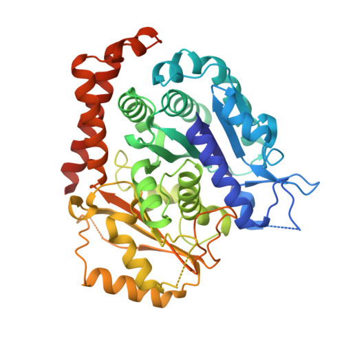
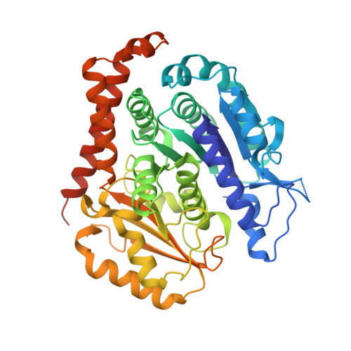
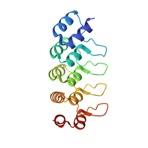
    7PJE, 7PJF

  • PubMed Abstract: 

    Infectious diseases caused by apicomplexan parasites remain a global public health threat. The presence of multiple ligand-binding sites in tubulin makes this protein an attractive target for anti-parasite drug discovery. However, despite remarkable successes as anti-cancer agents, the rational development of protozoan parasite-specific tubulin drugs has been hindered by a lack of structural and biochemical information on protozoan tubulins. Here, we present atomic structures for a protozoan tubulin and microtubule and delineate the architectures of apicomplexan tubulin drug-binding sites. Based on this information, we rationally designed the parasite-specific tubulin inhibitor parabulin and show that it inhibits growth of parasites while displaying no effects on human cells. Our work presents for the first time the rational design of a species-specific tubulin drug providing a framework to exploit structural differences between human and protozoa tubulin variants enabling the development of much-needed, novel parasite inhibitors.


  • Organizational Affiliation
    • Laboratory of Biomolecular Research, Division of Biology and Chemistry, Paul Scherrer Institut, Villigen, Switzerland.

Macromolecule Content 

  • Total Structure Weight: 118.44 kDa 
  • Atom Count: 8,262 
  • Modeled Residue Count: 1,002 
  • Deposited Residue Count: 1,058 
  • Unique protein chains: 3

Macromolecules

Find similar proteins by:|  3D Structure
Entity ID: 1
MoleculeChains  Sequence LengthOrganismDetailsImage
Tubulin alpha-1B chain451Homo sapiensMutation(s): 0 
Gene Names: TUBA1B
EC: 3.6.5
UniProt & NIH Common Fund Data Resources
Find proteins for P68363 (Homo sapiens)
Explore P68363 
Go to UniProtKB:  P68363
PHAROS:  P68363
GTEx:  ENSG00000123416 
Entity Groups
Sequence Clusters30% Identity50% Identity70% Identity90% Identity95% Identity100% Identity
UniProt GroupP68363
Sequence Annotations
Expand
Reference Sequence
Find similar proteins by:|  3D Structure
Entity ID: 2
MoleculeChains  Sequence LengthOrganismDetailsImage
Tubulin beta-3 chain450Homo sapiensMutation(s): 0 
Gene Names: TUBB3TUBB4
UniProt & NIH Common Fund Data Resources
Find proteins for Q13509 (Homo sapiens)
Explore Q13509 
Go to UniProtKB:  Q13509
PHAROS:  Q13509
GTEx:  ENSG00000258947 
Entity Groups
Sequence Clusters30% Identity50% Identity70% Identity90% Identity95% Identity100% Identity
UniProt GroupQ13509
Sequence Annotations
Expand
Reference Sequence
Find similar proteins by:|  3D Structure
Entity ID: 3
MoleculeChains  Sequence LengthOrganismDetailsImage
Designed ankyrin repeat protein (DARPIN) D1C [auth F]157synthetic constructMutation(s): 0 

Experimental Data & Validation

Experimental Data

  • Method: X-RAY DIFFRACTION
  • Resolution: 1.86 Å
  • R-Value Free:  0.218 (Depositor), 0.217 (DCC) 
  • R-Value Work:  0.184 (Depositor), 0.182 (DCC) 
  • R-Value Observed: 0.185 (Depositor) 
Space Group: P 1 21 1
Unit Cell:
Length ( Å )Angle ( ˚ )
a = 73.784α = 90
b = 91.312β = 97.55
c = 82.657γ = 90
Software Package:
Software NamePurpose
PHENIXrefinement
PDB_EXTRACTdata extraction
XDSdata reduction
XSCALEdata scaling
PHASERphasing

Structure Validation

View Full Validation Report


Ligand Structure Quality Assessment 


Entry History 

& Funding Information

Deposition Data


Funding OrganizationLocationGrant Number
Novartis FreeNovationSwitzerland--

Revision History  (Full details and data files)

  • Version 1.0: 2021-09-22
    Type: Initial release
  • Version 1.1: 2021-11-03
    Changes: Data collection, Database references
  • Version 1.2: 2021-11-17
    Changes: Data collection, Database references
  • Version 1.3: 2024-01-31
    Changes: Data collection, Refinement description