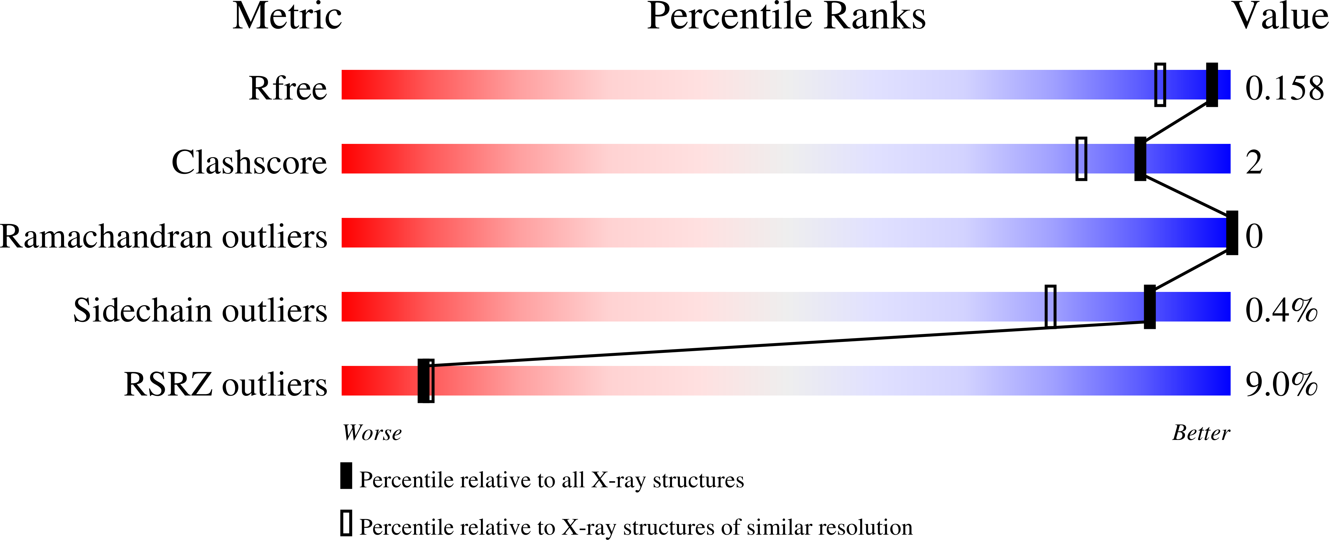
7PIH

Protein kinase A catalytic subunit in complex with PKI5-24 and EN093


Experimental Data Snapshot

  • Method: X-RAY DIFFRACTION
  • Resolution: 1.37 Å
  • R-Value Free: 0.157 
  • R-Value Work: 0.139 
  • R-Value Observed: 0.140 

Starting Model: experimental
View more details

wwPDB Validation   3D Report Full Report


Ligand Structure Quality Assessment 

Created with Raphaël 2.3.0Worse 01 BetterLigand structure goodness of fit to experimental dataBest fitted 7QWClick on this verticalbar to view details

This is version 1.2 of the entry. See complete history


Literature

Magnet for the Needle in Haystack: "Crystal Structure First" Fragment Hits Unlock Active Chemical Matter Using Targeted Exploration of Vast Chemical Spaces.

Muller, J.Klein, R.Tarkhanova, O.Gryniukova, A.Borysko, P.Merkl, S.Ruf, M.Neumann, A.Gastreich, M.Moroz, Y.S.Klebe, G.Glinca, S.

(2022) J Med Chem 65: 15663-15678

  • DOI: https://doi.org/10.1021/acs.jmedchem.2c00813
  • Primary Citation of Related Structures:  
    7PID, 7PIE, 7PIF, 7PIG, 7PIH, 7PNS

  • PubMed Abstract: 

    Fragment-based drug discovery (FBDD) has successfully led to approved therapeutics for challenging and "undruggable" targets. In the context of FBDD, we introduce a novel, multidisciplinary method to identify active molecules from purchasable chemical space. Starting from four small-molecule fragment complexes of protein kinase A (PKA), a template-based docking screen using Enamine's multibillion REAL Space was performed. A total of 93 molecules out of 106 selected compounds were successfully synthesized. Forty compounds were active in at least one validation assay with the most active follow-up having a 13,500-fold gain in affinity. Crystal structures for six of the most promising binders were rapidly obtained, verifying the binding mode. The overall success rate for this novel fragment-to-hit approach was 40%, accomplished in only 9 weeks. The results challenge the established fragment prescreening paradigm since the standard industrial filters for fragment hit identification in a thermal shift assay would have missed the initial fragments.


  • Organizational Affiliation

    CrystalsFirst GmbH, Marbacher Weg 6, 35037Marburg, Germany.


Macromolecules
Find similar proteins by:  (by identity cutoff)  |  3D Structure
Entity ID: 1
MoleculeChains Sequence LengthOrganismDetailsImage
cAMP-dependent protein kinase catalytic subunit alpha353Cricetulus griseusMutation(s): 0 
Gene Names: PRKACA
EC: 2.7.11.11
UniProt
Find proteins for P25321 (Cricetulus griseus)
Explore P25321 
Go to UniProtKB:  P25321
Entity Groups  
Sequence Clusters30% Identity50% Identity70% Identity90% Identity95% Identity100% Identity
UniProt GroupP25321
Sequence Annotations
Expand
  • Reference Sequence

Find similar proteins by:  Sequence   |   3D Structure  

Entity ID: 2
MoleculeChains Sequence LengthOrganismDetailsImage
cAMP-dependent protein kinase inhibitor alpha20Mus musculusMutation(s): 0 
UniProt & NIH Common Fund Data Resources
Find proteins for P63248 (Mus musculus)
Explore P63248 
Go to UniProtKB:  P63248
IMPC:  MGI:104747
Entity Groups  
Sequence Clusters30% Identity50% Identity70% Identity90% Identity95% Identity100% Identity
UniProt GroupP63248
Sequence Annotations
Expand
  • Reference Sequence
Small Molecules
Ligands 3 Unique
IDChains Name / Formula / InChI Key2D Diagram3D Interactions
7QW (Subject of Investigation/LOI)
Query on 7QW

Download Ideal Coordinates CCD File 
C [auth A]6-[3-[[(3~{R})-3-azanylpyrrolidin-1-yl]methyl]phenyl]-4~{H}-isoquinolin-1-one
C20 H21 N3 O
KJCKVXKGXLPESD-GOSISDBHSA-N
MPD
Query on MPD

Download Ideal Coordinates CCD File 
D [auth A](4S)-2-METHYL-2,4-PENTANEDIOL
C6 H14 O2
SVTBMSDMJJWYQN-YFKPBYRVSA-N
CL
Query on CL

Download Ideal Coordinates CCD File 
E [auth A]CHLORIDE ION
Cl
VEXZGXHMUGYJMC-UHFFFAOYSA-M
Modified Residues  2 Unique
IDChains TypeFormula2D DiagramParent
SEP
Query on SEP
A
L-PEPTIDE LINKINGC3 H8 N O6 PSER
TPO
Query on TPO
A
L-PEPTIDE LINKINGC4 H10 N O6 PTHR
Experimental Data & Validation

Experimental Data

  • Method: X-RAY DIFFRACTION
  • Resolution: 1.37 Å
  • R-Value Free: 0.157 
  • R-Value Work: 0.139 
  • R-Value Observed: 0.140 
  • Space Group: P 21 21 21
Unit Cell:
Length ( Å )Angle ( ˚ )
a = 58.49α = 90
b = 73.255β = 90
c = 109.029γ = 90
Software Package:
Software NamePurpose
PHENIXrefinement
XDSdata reduction
XDSdata scaling
PHASERphasing

Structure Validation

View Full Validation Report


Ligand Structure Quality Assessment 

Created with Raphaël 2.3.0Worse 01 BetterLigand structure goodness of fit to experimental dataBest fitted 7QWClick on this verticalbar to view details

Entry History & Funding Information

Deposition Data


Funding OrganizationLocationGrant Number
Not funded--

Revision History  (Full details and data files)

  • Version 1.0: 2022-09-21
    Type: Initial release
  • Version 1.1: 2022-12-21
    Changes: Database references
  • Version 1.2: 2024-01-31
    Changes: Data collection, Refinement description