7PD0 | pdb_00007pd0

Functional and structural characterization of redox sensitive superfolder green fluorescent protein and variants


Experimental Data Snapshot

  • Method: X-RAY DIFFRACTION
  • Resolution: 2.00 Å
  • R-Value Free: 
    0.298 (Depositor), 0.298 (DCC) 
  • R-Value Work: 
    0.250 (Depositor), 0.250 (DCC) 
  • R-Value Observed: 
    0.253 (Depositor) 

Starting Model: experimental
View more details

wwPDB Validation 3D Report Full Report

Validation slider image for 7PD0

This is version 1.3 of the entry. See complete history

Literature

Structure and Function of Redox-Sensitive Superfolder Green Fluorescent Protein Variant.

Heimsch, K.C.Gertzen, C.G.W.Schuh, A.K.Nietzel, T.Rahlfs, S.Przyborski, J.M.Gohlke, H.Schwarzlander, M.Becker, K.Fritz-Wolf, K.

(2022) Antioxid Redox Signal 37: 1-18

  • DOI: https://doi.org/10.1089/ars.2021.0234
  • Primary Citation Related Structures: 
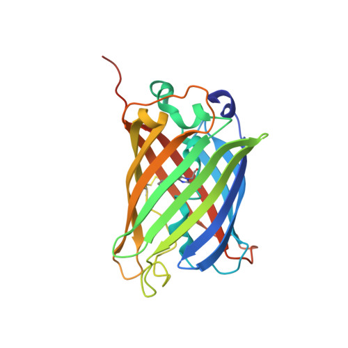
    7PCA, 7PCZ, 7PD0

  • PubMed Abstract: 

    Aims: Genetically encoded green fluorescent protein (GFP)-based redox biosensors are widely used to monitor specific and dynamic redox processes in living cells. Over the last few years, various biosensors for a variety of applications were engineered and enhanced to match the organism and cellular environments, which should be investigated. In this context, the unicellular intraerythrocytic parasite Plasmodium , the causative agent of malaria, represents a challenge, as the small size of the organism results in weak fluorescence signals that complicate precise measurements, especially for cell compartment-specific observations. To address this, we have functionally and structurally characterized an enhanced redox biosensor superfolder roGFP2 (sfroGFP2). Results: SfroGFP2 retains roGFP2-like behavior, yet with improved fluorescence intensity (FI) in cellulo . SfroGFP2-based redox biosensors are pH insensitive in a physiological pH range and show midpoint potentials comparable with roGFP2-based redox biosensors. Using crystallography and rigidity theory, we identified the superfolding mutations as being responsible for improved structural stability of the biosensor in a redox-sensitive environment, thus explaining the improved FI in cellulo . Innovation: This work provides insight into the structure and function of GFP-based redox biosensors. It describes an improved redox biosensor (sfroGFP2) suitable for measuring oxidizing effects within small cells where applicability of other redox sensor variants is limited. Conclusion: Improved structural stability of sfroGFP2 gives rise to increased FI in cellulo . Fusion to hGrx1 (human glutaredoxin-1) provides the hitherto most suitable biosensor for measuring oxidizing effects in Plasmodium . This sensor is of major interest for studying glutathione redox changes in small cells, as well as subcellular compartments in general. Antioxid. Redox Signal. 37, 1-18.


  • Organizational Affiliation
    • Biochemistry and Molecular Biology, Interdisciplinary Research Center, Justus Liebig University Giessen, Giessen, Germany.

Macromolecule Content 

  • Total Structure Weight: 108.49 kDa 
  • Atom Count: 8,017 
  • Modeled Residue Count: 927 
  • Deposited Residue Count: 944 
  • Unique protein chains: 1

Macromolecules

Find similar proteins by:|  3D Structure
Entity ID: 1
MoleculeChains  Sequence LengthOrganismDetailsImage
Green fluorescent protein
A, B, C, D
236Aequorea victoriaMutation(s): 16 
Gene Names: GFP
UniProt
Find proteins for P42212 (Aequorea victoria)
Explore P42212 
Go to UniProtKB:  P42212
Entity Groups
Sequence Clusters30% Identity50% Identity70% Identity90% Identity95% Identity100% Identity
UniProt GroupP42212
Sequence Annotations
Expand
Reference Sequence

Small Molecules

Ligands 3 Unique
IDChains Name / Formula / InChI Key2D Diagram3D Interactions
PGE

Query on PGE


Download:Ideal Coordinates CCD File
E [auth A],
G [auth A],
J [auth C]
TRIETHYLENE GLYCOL
C6 H14 O4
ZIBGPFATKBEMQZ-UHFFFAOYSA-N
PEG

Query on PEG


Download:Ideal Coordinates CCD File
F [auth A]
H [auth B]
I [auth B]
K [auth C]
L [auth C]
F [auth A],
H [auth B],
I [auth B],
K [auth C],
L [auth C],
M [auth C],
N [auth D]
DI(HYDROXYETHYL)ETHER
C4 H10 O3
MTHSVFCYNBDYFN-UHFFFAOYSA-N
GOL

Query on GOL


Download:Ideal Coordinates CCD File
O [auth D]GLYCEROL
C3 H8 O3
PEDCQBHIVMGVHV-UHFFFAOYSA-N
Modified Residues  1 Unique
IDChains TypeFormula2D DiagramParent
CRO
Query on CRO
A, B, C, D
L-PEPTIDE LINKINGC15 H17 N3 O5THR, TYR, GLY

Experimental Data & Validation

Experimental Data

  • Method: X-RAY DIFFRACTION
  • Resolution: 2.00 Å
  • R-Value Free:  0.298 (Depositor), 0.298 (DCC) 
  • R-Value Work:  0.250 (Depositor), 0.250 (DCC) 
  • R-Value Observed: 0.253 (Depositor) 
Space Group: P 1 21 1
Unit Cell:
Length ( Å )Angle ( ˚ )
a = 91.85α = 90
b = 51.29β = 102.28
c = 101.46γ = 90
Software Package:
Software NamePurpose
PHENIXrefinement
XDSdata reduction
XDSdata scaling
PHENIXphasing

Structure Validation

View Full Validation Report


Entry History 

& Funding Information

Deposition Data


Funding OrganizationLocationGrant Number
Hessian Ministry of Science, Higher Education and Art (HMWK)GermanyLoewe Druid E3

Revision History  (Full details and data files)

  • Version 1.0: 2022-02-16
    Type: Initial release
  • Version 1.1: 2022-07-20
    Changes: Database references
  • Version 1.2: 2024-01-31
    Changes: Data collection, Refinement description
  • Version 1.3: 2024-10-16
    Changes: Structure summary