7PA5 | pdb_00007pa5

Complex between the beta-lactamase CMY-2 with an inhibitory nanobody


Experimental Data Snapshot

  • Method: X-RAY DIFFRACTION
  • Resolution: 3.18 Å
  • R-Value Free: 
    0.250 (Depositor), 0.257 (DCC) 
  • R-Value Work: 
    0.207 (Depositor), 0.211 (DCC) 
  • R-Value Observed: 
    0.209 (Depositor) 

Starting Models: experimental
View more details

wwPDB Validation 3D Report Full Report

Validation slider image for 7PA5

This is version 1.2 of the entry. See complete history

Literature

Development of Nanobodies as Theranostic Agents against CMY-2-Like Class C beta-Lactamases.

Cawez, F.Mercuri, P.S.Morales-Yanez, F.J.Maalouf, R.Vandevenne, M.Kerff, F.Guerin, V.Mainil, J.Thiry, D.Saulmont, M.Vanderplasschen, A.Lafaye, P.Ayme, G.Bogaerts, P.Dumoulin, M.Galleni, M.

(2023) Antimicrob Agents Chemother 67: e0149922-e0149922

  • DOI: https://doi.org/10.1128/aac.01499-22
  • Primary Citation Related Structures: 
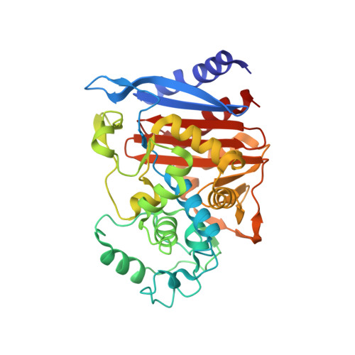
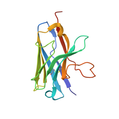
    7PA5

  • PubMed Abstract: 

    Three soluble single-domain fragments derived from the unique variable region of camelid heavy-chain antibodies (VHHs) against the CMY-2 β-lactamase behaved as inhibitors. The structure of the complex VHH cAb CMY-2 (254)/CMY-2 showed that the epitope is close to the active site and that the CDR3 of the VHH protrudes into the catalytic site. The β-lactamase inhibition pattern followed a mixed profile with a predominant noncompetitive component. The three isolated VHHs recognized overlapping epitopes since they behaved as competitive binders. Our study identified a binding site that can be targeted by a new class of β-lactamase inhibitors designed on the sequence of the paratope. Furthermore, the use of mono- or bivalent VHH and rabbit polyclonal anti-CMY-2 antibodies enables the development of the first generation of enzyme-linked immunosorbent assay (ELISA) for the detection of CMY-2 produced by CMY-2-expressing bacteria, irrespective of resistotype.


  • Organizational Affiliation
    • InBioS, Center for Protein Engineering, Biological Macromolecules, Department of Life Sciences, University of Liège, Liège, Belgium.

Macromolecule Content 

  • Total Structure Weight: 54.29 kDa 
  • Atom Count: 3,773 
  • Modeled Residue Count: 481 
  • Deposited Residue Count: 490 
  • Unique protein chains: 2

Macromolecules

Find similar proteins by:|  3D Structure
Entity ID: 1
MoleculeChains  Sequence LengthOrganismDetailsImage
Beta-lactamase361Escherichia coliMutation(s): 0 
Gene Names: 
EC: 3.5.2.6
UniProt
Find proteins for Q53TY8 (Escherichia coli)
Explore Q53TY8 
Go to UniProtKB:  Q53TY8
Entity Groups
Sequence Clusters30% Identity50% Identity70% Identity90% Identity95% Identity100% Identity
UniProt GroupQ53TY8
Sequence Annotations
Expand
Reference Sequence
Find similar proteins by:|  3D Structure
Entity ID: 2
MoleculeChains  Sequence LengthOrganismDetailsImage
nanobody129CamelidaeMutation(s): 0 

Experimental Data & Validation

Experimental Data

  • Method: X-RAY DIFFRACTION
  • Resolution: 3.18 Å
  • R-Value Free:  0.250 (Depositor), 0.257 (DCC) 
  • R-Value Work:  0.207 (Depositor), 0.211 (DCC) 
  • R-Value Observed: 0.209 (Depositor) 
Space Group: P 62 2 2
Unit Cell:
Length ( Å )Angle ( ˚ )
a = 95.12α = 90
b = 95.12β = 90
c = 242.94γ = 120
Software Package:
Software NamePurpose
XDSdata reduction
XSCALEdata scaling
PHASERphasing
PHENIXrefinement
PDB_EXTRACTdata extraction

Structure Validation

View Full Validation Report


Entry History 

Deposition Data

Revision History  (Full details and data files)

  • Version 1.0: 2022-11-16
    Type: Initial release
  • Version 1.1: 2024-01-31
    Changes: Data collection, Database references, Refinement description
  • Version 1.2: 2024-11-06
    Changes: Structure summary