7P84 | pdb_00007p84

Crystal structure of L147A/I351A variant of S-adenosylmethionine synthetase from Methanocaldococcus jannaschii in complex with ONB-SAM (2-nitro benzyme S-adenosyl-methionine)


Experimental Data Snapshot

  • Method: X-RAY DIFFRACTION
  • Resolution: 2.05 Å
  • R-Value Free: 
    0.222 (Depositor), 0.222 (DCC) 
  • R-Value Work: 
    0.178 (Depositor), 0.183 (DCC) 
  • R-Value Observed: 
    0.178 (Depositor) 

Starting Model: experimental
View more details

wwPDB Validation 3D Report Full Report

Validation slider image for 7P84

Ligand Structure Quality Assessment 


This is version 1.2 of the entry. See complete history

Literature

Visible-Light Removable Photocaging Groups Accepted by MjMAT Variant: Structural Basis and Compatibility with DNA and RNA Methyltransferases.

Peters, A.Herrmann, E.Cornelissen, N.V.Klocker, N.Kummel, D.Rentmeister, A.

(2022) Chembiochem 23: e202100437-e202100437

  • DOI: https://doi.org/10.1002/cbic.202100437
  • Primary Citation Related Structures: 
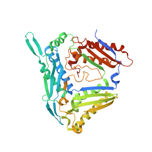
    7P82, 7P83, 7P84, 7P8M

  • PubMed Abstract: 

    Methylation and demethylation of DNA, RNA and proteins constitutes a major regulatory mechanism in epigenetic processes. Investigations would benefit from the ability to install photo-cleavable groups at methyltransferase target sites that block interactions with reader proteins until removed by non-damaging light in the visible spectrum. Engineered methionine adenosyltransferases (MATs) have been exploited in cascade reactions with methyltransferases (MTases) to modify biomolecules with non-natural groups, including first evidence for accepting photo-cleavable groups. We show that an engineered MAT from Methanocaldococcus jannaschii (PC-MjMAT) is 308-fold more efficient at converting ortho-nitrobenzyl-(ONB)-homocysteine than the wildtype enzyme. PC-MjMAT is active over a broad range of temperatures and compatible with MTases from mesophilic organisms. We solved the crystal structures of wildtype and PC-MjMAT in complex with AdoONB and a red-shifted derivative thereof. These structures reveal that aromatic stacking interactions within the ligands are key to accommodating the photocaging groups in PC-MjMAT. The enlargement of the binding pocket eliminates steric clashes to enable AdoMet analogue binding. Importantly, PC-MjMAT exhibits remarkable activity on methionine analogues with red-shifted ONB-derivatives enabling photo-deprotection of modified DNA by visible light.


  • Organizational Affiliation
    • Department of Chemistry and Pharmacy, Institute of Biochemistry, University of Münster, Corrensstr. 36, 48149, Münster, Germany.

Macromolecule Content 

  • Total Structure Weight: 95.91 kDa 
  • Atom Count: 6,798 
  • Modeled Residue Count: 814 
  • Deposited Residue Count: 852 
  • Unique protein chains: 1

Macromolecules

Find similar proteins by:|  3D Structure
Entity ID: 1
MoleculeChains  Sequence LengthOrganismDetailsImage
S-adenosylmethionine synthaseA,
B [auth C]
426Methanocaldococcus jannaschii DSM 2661Mutation(s): 2 
Gene Names: matMJ1208
EC: 2.5.1.6
UniProt
Find proteins for Q58605 (Methanocaldococcus jannaschii (strain ATCC 43067 / DSM 2661 / JAL-1 / JCM 10045 / NBRC 100440))
Explore Q58605 
Go to UniProtKB:  Q58605
Entity Groups
Sequence Clusters30% Identity50% Identity70% Identity90% Identity95% Identity100% Identity
UniProt GroupQ58605
Sequence Annotations
Expand
Reference Sequence

Small Molecules

Ligands 3 Unique
IDChains Name / Formula / InChI Key2D Diagram3D Interactions
EU9
(Subject of Investigation/LOI)

Query on EU9


Download:Ideal Coordinates CCD File
H [auth C][(2S,3S,4R,5R)-5-(6-aminopurin-9-yl)-3,4-bis(oxidanyl)oxolan-2-yl]methyl-[(3R)-3-azanyl-4-oxidanyl-4-oxidanylidene-butyl]-[(2-nitrophenyl)methyl]sulfanium
C21 H26 N7 O7 S
UTGYKEAAXXLVQJ-OUPAZNFWSA-O
3PO

Query on 3PO


Download:Ideal Coordinates CCD File
C [auth A],
F [auth C]
TRIPHOSPHATE
H5 O10 P3
UNXRWKVEANCORM-UHFFFAOYSA-N
MG

Query on MG


Download:Ideal Coordinates CCD File
D [auth A],
E [auth A],
G [auth C]
MAGNESIUM ION
Mg
JLVVSXFLKOJNIY-UHFFFAOYSA-N

Experimental Data & Validation

Experimental Data

  • Method: X-RAY DIFFRACTION
  • Resolution: 2.05 Å
  • R-Value Free:  0.222 (Depositor), 0.222 (DCC) 
  • R-Value Work:  0.178 (Depositor), 0.183 (DCC) 
  • R-Value Observed: 0.178 (Depositor) 
Space Group: P 32 2 1
Unit Cell:
Length ( Å )Angle ( ˚ )
a = 76.104α = 90
b = 76.104β = 90
c = 280.674γ = 120
Software Package:
Software NamePurpose
PHENIXrefinement
PDB_EXTRACTdata extraction
XDSdata reduction
XSCALEdata scaling
PHASERphasing

Structure Validation

View Full Validation Report


Ligand Structure Quality Assessment 


Entry History 

Deposition Data

Revision History  (Full details and data files)

  • Version 1.0: 2021-11-17
    Type: Initial release
  • Version 1.1: 2022-01-19
    Changes: Database references
  • Version 1.2: 2024-01-31
    Changes: Data collection, Refinement description