7P7O | pdb_00007p7o

X-RAY CRYSTAL STRUCTURE OF SPOROSARCINA PASTEURII UREASE INHIBITED BY THE GOLD(I)-DIPHOSPHINE COMPOUND Au(PEt3)2Cl DETERMINED AT 1.87 ANGSTROMS


Experimental Data Snapshot

  • Method: X-RAY DIFFRACTION
  • Resolution: 1.87 Å
  • R-Value Free: 
    0.210 (Depositor), 0.212 (DCC) 
  • R-Value Work: 
    0.165 (Depositor), 0.167 (DCC) 

Starting Model: experimental
View more details

wwPDB Validation 3D Report Full Report

Validation slider image for 7P7O

Ligand Structure Quality Assessment 


This is version 1.1 of the entry. See complete history

Literature

Medicinal Au(I) compounds targeting urease as prospective antimicrobial agents: unveiling the structural basis for enzyme inhibition.

Mazzei, L.Massai, L.Cianci, M.Messori, L.Ciurli, S.

(2021) Dalton Trans 50: 14444-14452

  • DOI: https://doi.org/10.1039/d1dt02488d
  • Primary Citation Related Structures: 
    7P7N, 7P7O

  • PubMed Abstract: 

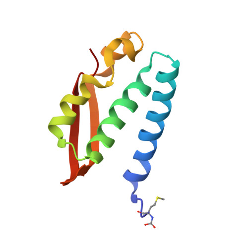
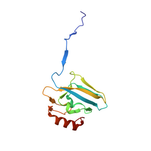
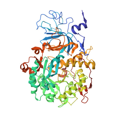
    A few gold compounds were recently found to show antimicrobial properties in vitro , holding great promise for the discovery of new drugs to overcome antibiotic resistance. Here, the inhibition of the bacterial virulence factor urease by four Au(I)-compounds, namely Au(PEt 3 )Cl, Au(PEt 3 )Br, Au(PEt 3 )I and [Au(PEt 3 ) 2 ]Cl, obtained from the antiarthritic Au(I)-drug Auranofin and earlier reported to act as antimicrobials, is investigated. The three monophosphino Au(I) complexes showed IC 50 values in the 30-100 nM range, while the diphosphino Au(I) complex, though being less active, still showed a IC 50 value of 7 μM. The structural basis for this inhibition was provided by solving the crystal structures of urease co-crystallized with Au(PEt 3 )I and [Au(PEt 3 ) 2 ]Cl: at least two Au(I) ions bind the enzyme in a flap domain involved in the catalysis, thus obliterating enzyme activity. Peculiar changes observed in the two structures reveal implications for the mechanism of soft metal binding and enzyme inactivation.


  • Organizational Affiliation
    • Laboratory of Bioinorganic Chemistry, Department of Pharmacy and Biotechnology (FaBiT), University of Bologna, Via Giuseppe Fanin 40, I-40127 Bologna, Italy. luca.mazzei2@unibo.it.

Macromolecule Content 

  • Total Structure Weight: 89.58 kDa 
  • Atom Count: 6,880 
  • Modeled Residue Count: 785 
  • Deposited Residue Count: 792 
  • Unique protein chains: 3

Macromolecules

Find similar proteins by:|  3D Structure
Entity ID: 1
MoleculeChains  Sequence LengthOrganismDetailsImage
Urease subunit gammaA [auth AAA]100Sporosarcina pasteuriiMutation(s): 2 
EC: 3.5.1.5
UniProt
Find proteins for P41022 (Sporosarcina pasteurii)
Explore P41022 
Go to UniProtKB:  P41022
Entity Groups
Sequence Clusters30% Identity50% Identity70% Identity90% Identity95% Identity100% Identity
UniProt GroupP41022
Sequence Annotations
Expand
Reference Sequence
Find similar proteins by:|  3D Structure
Entity ID: 2
MoleculeChains  Sequence LengthOrganismDetailsImage
Urease subunit betaB [auth BBB]122Sporosarcina pasteuriiMutation(s): 0 
EC: 3.5.1.5
UniProt
Find proteins for P41021 (Sporosarcina pasteurii)
Explore P41021 
Go to UniProtKB:  P41021
Entity Groups
Sequence Clusters30% Identity50% Identity70% Identity90% Identity95% Identity100% Identity
UniProt GroupP41021
Sequence Annotations
Expand
Reference Sequence
Find similar proteins by:|  3D Structure
Entity ID: 3
MoleculeChains  Sequence LengthOrganismDetailsImage
Urease subunit alphaC [auth CCC]570Sporosarcina pasteuriiMutation(s): 0 
EC: 3.5.1.5
UniProt
Find proteins for P41020 (Sporosarcina pasteurii)
Explore P41020 
Go to UniProtKB:  P41020
Entity Groups
Sequence Clusters30% Identity50% Identity70% Identity90% Identity95% Identity100% Identity
UniProt GroupP41020
Sequence Annotations
Expand
Reference Sequence

Small Molecules

Ligands 5 Unique
IDChains Name / Formula / InChI Key2D Diagram3D Interactions
AUF

Query on AUF


Download:Ideal Coordinates CCD File
T [auth CCC],
U [auth CCC],
V [auth CCC]
triethylphosphanuidylgold(1+)
C6 H15 Au P
NKNLIDCIHVTIMI-UHFFFAOYSA-N
SO4

Query on SO4


Download:Ideal Coordinates CCD File
AA [auth CCC]
BA [auth CCC]
CA [auth CCC]
DA [auth CCC]
EA [auth CCC]
AA [auth CCC],
BA [auth CCC],
CA [auth CCC],
DA [auth CCC],
EA [auth CCC],
F [auth AAA],
FA [auth CCC],
G [auth AAA],
GA [auth CCC],
H [auth AAA],
HA [auth CCC],
I [auth AAA],
IA [auth CCC],
K [auth BBB],
L [auth BBB],
W [auth CCC],
X [auth CCC],
Y [auth CCC],
Z [auth CCC]
SULFATE ION
O4 S
QAOWNCQODCNURD-UHFFFAOYSA-L
EDO

Query on EDO


Download:Ideal Coordinates CCD File
D [auth AAA]
E [auth AAA]
J [auth BBB]
O [auth CCC]
Q [auth CCC]
D [auth AAA],
E [auth AAA],
J [auth BBB],
O [auth CCC],
Q [auth CCC],
R [auth CCC],
S [auth CCC]
1,2-ETHANEDIOL
C2 H6 O2
LYCAIKOWRPUZTN-UHFFFAOYSA-N
NI

Query on NI


Download:Ideal Coordinates CCD File
M [auth CCC],
N [auth CCC]
NICKEL (II) ION
Ni
VEQPNABPJHWNSG-UHFFFAOYSA-N
O

Query on O


Download:Ideal Coordinates CCD File
P [auth CCC]OXYGEN ATOM
O
XLYOFNOQVPJJNP-UHFFFAOYSA-N
Modified Residues  2 Unique
IDChains TypeFormula2D DiagramParent
CXM
Query on CXM
A [auth AAA]L-PEPTIDE LINKINGC6 H11 N O4 SMET
KCX
Query on KCX
C [auth CCC]L-PEPTIDE LINKINGC7 H14 N2 O4LYS

Experimental Data & Validation

Experimental Data

  • Method: X-RAY DIFFRACTION
  • Resolution: 1.87 Å
  • R-Value Free:  0.210 (Depositor), 0.212 (DCC) 
  • R-Value Work:  0.165 (Depositor), 0.167 (DCC) 
Space Group: P 63 2 2
Unit Cell:
Length ( Å )Angle ( ˚ )
a = 131.797α = 90
b = 131.797β = 90
c = 189.381γ = 120
Software Package:
Software NamePurpose
REFMACrefinement
XDSdata reduction
Aimlessdata scaling
REFMACphasing

Structure Validation

View Full Validation Report


Ligand Structure Quality Assessment 


Entry History 

& Funding Information

Deposition Data


Funding OrganizationLocationGrant Number
Not funded--

Revision History  (Full details and data files)

  • Version 1.0: 2022-06-01
    Type: Initial release
  • Version 1.1: 2024-01-31
    Changes: Data collection, Derived calculations, Refinement description