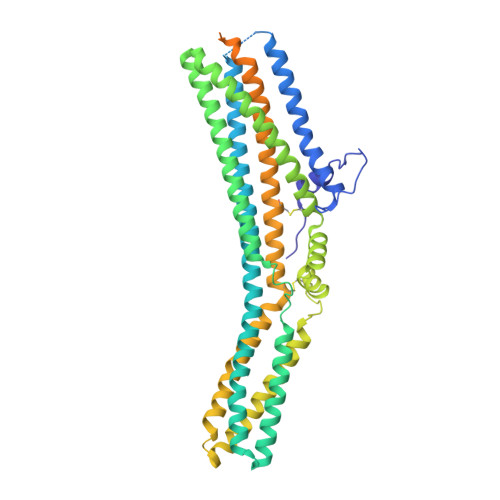
7P5C

Cryo-EM structure of human TTYH3 in Ca2+ and GDN


Experimental Data Snapshot

  • Method: ELECTRON MICROSCOPY
  • Resolution: 3.20 Å
  • Aggregation State: PARTICLE 
  • Reconstruction Method: SINGLE PARTICLE 

wwPDB Validation   3D Report Full Report


This is version 1.2 of the entry. See complete history


Literature

Cryo-EM structures of the TTYH family reveal a novel architecture for lipid interactions.

Sukalskaia, A.Straub, M.S.Deneka, D.Sawicka, M.Dutzler, R.

(2021) Nat Commun 12: 4893-4893

  • DOI: https://doi.org/10.1038/s41467-021-25106-4
  • Primary Citation of Related Structures:  
    7P54, 7P5C, 7P5J, 7P5M

  • PubMed Abstract: 

    The Tweety homologs (TTYHs) are members of a conserved family of eukaryotic membrane proteins that are abundant in the brain. The three human paralogs were assigned to function as anion channels that are either activated by Ca 2+ or cell swelling. To uncover their unknown architecture and its relationship to function, we have determined the structures of human TTYH1-3 by cryo-electron microscopy. All structures display equivalent features of a dimeric membrane protein that contains five transmembrane segments and an extended extracellular domain. As none of the proteins shows attributes reminiscent of an anion channel, we revisited functional experiments and did not find any indication of ion conduction. Instead, we find density in an extended hydrophobic pocket contained in the extracellular domain that emerges from the lipid bilayer, which suggests a role of TTYH proteins in the interaction with lipid-like compounds residing in the membrane.


  • Organizational Affiliation

    Department of Biochemistry, University of Zurich, Zurich, Switzerland.


Macromolecules
Find similar proteins by:  (by identity cutoff)  |  3D Structure
Entity ID: 1
MoleculeChains Sequence LengthOrganismDetailsImage
Protein tweety homolog 3
A, B
530Homo sapiensMutation(s): 0 
Gene Names: TTYH3KIAA1691
Membrane Entity: Yes 
UniProt & NIH Common Fund Data Resources
Find proteins for Q9C0H2 (Homo sapiens)
Explore Q9C0H2 
Go to UniProtKB:  Q9C0H2
PHAROS:  Q9C0H2
GTEx:  ENSG00000136295 
Entity Groups  
Sequence Clusters30% Identity50% Identity70% Identity90% Identity95% Identity100% Identity
UniProt GroupQ9C0H2
Glycosylation
Glycosylation Sites: 3Go to GlyGen: Q9C0H2-1
Sequence Annotations
Expand
  • Reference Sequence
Oligosaccharides

Help

Entity ID: 2
MoleculeChains Length2D Diagram Glycosylation3D Interactions
2-acetamido-2-deoxy-beta-D-glucopyranose-(1-4)-2-acetamido-2-deoxy-beta-D-glucopyranose
C, D
2N-Glycosylation
Glycosylation Resources
GlyTouCan:  G42666HT
GlyCosmos:  G42666HT
GlyGen:  G42666HT
Experimental Data & Validation

Experimental Data

  • Method: ELECTRON MICROSCOPY
  • Resolution: 3.20 Å
  • Aggregation State: PARTICLE 
  • Reconstruction Method: SINGLE PARTICLE 

Structure Validation

View Full Validation Report


Entry History & Funding Information

Deposition Data


Funding OrganizationLocationGrant Number
Swiss National Science FoundationSwitzerland31003A_163421

Revision History  (Full details and data files)

  • Version 1.0: 2021-08-11
    Type: Initial release
  • Version 1.1: 2021-08-25
    Changes: Database references, Structure summary
  • Version 1.2: 2021-09-08
    Changes: Database references