7OYV | pdb_00007oyv

E.coli's putrescine receptor variant PotF/D (4JDF) with mutations E39D F88A S247D in complex with spermidine


Experimental Data Snapshot

  • Method: X-RAY DIFFRACTION
  • Resolution: 1.90 Å
  • R-Value Free: 
    0.276 (Depositor), 0.273 (DCC) 
  • R-Value Work: 
    0.227 (Depositor), 0.227 (DCC) 
  • R-Value Observed: 
    0.229 (Depositor) 

Starting Model: experimental
View more details

wwPDB Validation 3D Report Full Report

Validation slider image for 7OYV

Ligand Structure Quality Assessment 


This is version 1.3 of the entry. See complete history

Literature

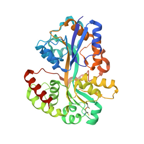
Fine-tuning spermidine binding modes in the putrescine binding protein PotF.

Kroger, P.Shanmugaratnam, S.Scheib, U.Hocker, B.

(2021) J Biological Chem 297: 101419-101419

  • DOI: https://doi.org/10.1016/j.jbc.2021.101419
  • Primary Citation Related Structures: 
    7OYS, 7OYT, 7OYU, 7OYV, 7OYW, 7OYX, 7OYY, 7OYZ

  • PubMed Abstract: 

    A profound understanding of the molecular interactions between receptors and ligands is important throughout diverse research, such as protein design, drug discovery, or neuroscience. What determines specificity and how do proteins discriminate against similar ligands? In this study, we analyzed factors that determine binding in two homologs belonging to the well-known superfamily of periplasmic binding proteins, PotF and PotD. Building on a previously designed construct, modes of polyamine binding were swapped. This change of specificity was approached by analyzing local differences in the binding pocket as well as overall conformational changes in the protein. Throughout the study, protein variants were generated and characterized structurally and thermodynamically, leading to a specificity swap and improvement in affinity. This dataset not only enriches our knowledge applicable to rational protein design but also our results can further lay groundwork for engineering of specific biosensors as well as help to explain the adaptability of pathogenic bacteria.


  • Organizational Affiliation
    • Department for Biochemistry, University of Bayreuth, Bayreuth, Germany.

Macromolecule Content 

  • Total Structure Weight: 80.29 kDa 
  • Atom Count: 5,749 
  • Modeled Residue Count: 682 
  • Deposited Residue Count: 704 
  • Unique protein chains: 1

Macromolecules

Find similar proteins by:|  3D Structure
Entity ID: 1
MoleculeChains  Sequence LengthOrganismDetailsImage
Putrescine-binding periplasmic protein
A, B
352Escherichia coliMutation(s): 6 
Gene Names: potFHPE39_07830WP7S17E04_28980
UniProt
Find proteins for P31133 (Escherichia coli (strain K12))
Explore P31133 
Go to UniProtKB:  P31133
Entity Groups
Sequence Clusters30% Identity50% Identity70% Identity90% Identity95% Identity100% Identity
UniProt GroupP31133
Sequence Annotations
Expand
Reference Sequence

Small Molecules

Ligands 5 Unique
IDChains Name / Formula / InChI Key2D Diagram3D Interactions
3IZ

Query on 3IZ


Download:Ideal Coordinates CCD File
C [auth A](2~{R})-1-[(2~{R})-1-[(2~{R})-1-(2-methoxyethoxy)propan-2-yl]oxypropan-2-yl]oxypropan-2-amine
C12 H27 N O4
JAVLCTSZYWYEEE-IJLUTSLNSA-N
SPD
(Subject of Investigation/LOI)

Query on SPD


Download:Ideal Coordinates CCD File
F [auth A],
M [auth B]
SPERMIDINE
C7 H19 N3
ATHGHQPFGPMSJY-UHFFFAOYSA-N
3IE

Query on 3IE


Download:Ideal Coordinates CCD File
G [auth A],
H [auth A],
N [auth B]
(2~{S})-1-methoxypropan-2-amine
C4 H11 N O
NXMXETCTWNXSFG-BYPYZUCNSA-N
JFN

Query on JFN


Download:Ideal Coordinates CCD File
D [auth A],
E [auth A],
J [auth B],
K [auth B],
L [auth B]
(2R)-1-methoxypropan-2-amine
C4 H11 N O
NXMXETCTWNXSFG-SCSAIBSYSA-N
CL

Query on CL


Download:Ideal Coordinates CCD File
I [auth A],
O [auth B]
CHLORIDE ION
Cl
VEXZGXHMUGYJMC-UHFFFAOYSA-M

Experimental Data & Validation

Experimental Data

  • Method: X-RAY DIFFRACTION
  • Resolution: 1.90 Å
  • R-Value Free:  0.276 (Depositor), 0.273 (DCC) 
  • R-Value Work:  0.227 (Depositor), 0.227 (DCC) 
  • R-Value Observed: 0.229 (Depositor) 
Space Group: C 2 2 21
Unit Cell:
Length ( Å )Angle ( ˚ )
a = 73.613α = 90
b = 122.576β = 90
c = 190.32γ = 90
Software Package:
Software NamePurpose
PHENIXrefinement
Cootmodel building
XDSdata reduction
XSCALEdata scaling
PHASERphasing

Structure Validation

View Full Validation Report


Ligand Structure Quality Assessment 


Entry History 

& Funding Information

Deposition Data


Funding OrganizationLocationGrant Number
German Research Foundation (DFG)GermanyHO4022/2-3

Revision History  (Full details and data files)

  • Version 1.0: 2021-12-01
    Type: Initial release
  • Version 1.1: 2021-12-22
    Changes: Database references
  • Version 1.2: 2024-01-31
    Changes: Data collection, Refinement description
  • Version 1.3: 2024-11-20
    Changes: Structure summary