7OVO | pdb_00007ovo

Heterodimeric murine tRNA-guanine transglycosylase in complex with queuine


Experimental Data Snapshot

  • Method: X-RAY DIFFRACTION
  • Resolution: 2.10 Å
  • R-Value Free: 
    0.234 (Depositor), 0.234 (DCC) 
  • R-Value Work: 
    0.189 (Depositor), 0.192 (DCC) 
  • R-Value Observed: 
    0.191 (Depositor) 

Starting Models: experimental
View more details

wwPDB Validation 3D Report Full Report

Validation slider image for 7OVO

Ligand Structure Quality Assessment 


This is version 1.2 of the entry. See complete history

Literature

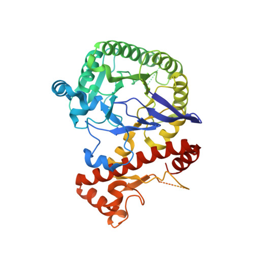
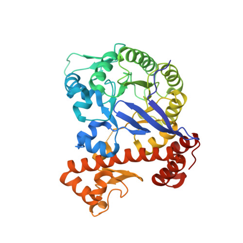
Structural and Biochemical Investigation of the Heterodimeric Murine tRNA-Guanine Transglycosylase.

Sebastiani, M.Behrens, C.Dorr, S.Gerber, H.D.Benazza, R.Hernandez-Alba, O.Cianferani, S.Klebe, G.Heine, A.Reuter, K.

(2022) ACS Chem Biol 17: 2229-2247

  • DOI: https://doi.org/10.1021/acschembio.2c00368
  • Primary Citation Related Structures: 
    6H62, 7B2I, 7OV9, 7OVO, 7OVS, 7OWZ

  • PubMed Abstract: 

    In tRNA Asp , tRNA Asn , tRNA Tyr , and tRNA His of most bacteria and eukaryotes, the anticodon wobble position may be occupied by the modified nucleoside queuosine, which affects the speed and the accuracy of translation. Since eukaryotes are not able to synthesize queuosine de novo, they have to salvage queuine (the queuosine base) as a micronutrient from food and/or the gut microbiome. The heterodimeric Zn 2+ containing enzyme tRNA-guanine transglycosylase (TGT) catalyzes the insertion of queuine into the above-named tRNAs in exchange for the genetically encoded guanine. This enzyme has attracted medical interest since it was shown to be potentially useful for the treatment of multiple sclerosis. In addition, TGT inactivation via gene knockout leads to the suppressed cell proliferation and migration of certain breast cancer cells, which may render this enzyme a potential target for the design of compounds supporting breast cancer therapy. As a prerequisite to fully exploit the medical potential of eukaryotic TGT, we have determined and analyzed a number of crystal structures of the functional murine TGT with and without bound queuine. In addition, we have investigated the importance of two residues of its non-catalytic subunit on dimer stability and determined the Michaelis-Menten parameters of murine TGT with respect to tRNA and several natural and artificial nucleobase substrates. Ultimately, on the basis of available TGT crystal structures, we provide an entirely conclusive reaction mechanism for this enzyme, which in detail explains why the TGT-catalyzed insertion of some nucleobases into tRNA occurs reversibly while that of others is irreversible.


  • Organizational Affiliation
    • Institut für Pharmazeutische Chemie, Philipps-Universität Marburg, Marbacher Weg 8, D-35037 Marburg, Germany.

Macromolecule Content 

  • Total Structure Weight: 91.3 kDa 
  • Atom Count: 5,997 
  • Modeled Residue Count: 755 
  • Deposited Residue Count: 814 
  • Unique protein chains: 2

Macromolecules

Find similar proteins by:|  3D Structure
Entity ID: 1
MoleculeChains  Sequence LengthOrganismDetailsImage
Queuine tRNA-ribosyltransferase accessory subunit 2419Mus musculusMutation(s): 0 
Gene Names: Qtrt2Qtrtd1
EC: 2.4.2.29
UniProt
Find proteins for B8ZXI1 (Mus musculus)
Explore B8ZXI1 
Go to UniProtKB:  B8ZXI1
Entity Groups
Sequence Clusters30% Identity50% Identity70% Identity90% Identity95% Identity100% Identity
UniProt GroupB8ZXI1
Sequence Annotations
Expand
Reference Sequence
Find similar proteins by:|  3D Structure
Entity ID: 2
MoleculeChains  Sequence LengthOrganismDetailsImage
Queuine tRNA-ribosyltransferase catalytic subunit 1B [auth C]395Mus musculusMutation(s): 0 
Gene Names: Qtrt1TgtTgut
EC: 2.4.2.29 (PDB Primary Data), 2.4.2.64 (UniProt)
UniProt
Find proteins for Q9JMA2 (Mus musculus)
Explore Q9JMA2 
Go to UniProtKB:  Q9JMA2
Entity Groups
Sequence Clusters30% Identity50% Identity70% Identity90% Identity95% Identity100% Identity
UniProt GroupQ9JMA2
Sequence Annotations
Expand
Reference Sequence

Small Molecules

Ligands 4 Unique
IDChains Name / Formula / InChI Key2D Diagram3D Interactions
QEI
(Subject of Investigation/LOI)

Query on QEI


Download:Ideal Coordinates CCD File
G [auth C]2-amino-5-({[(1S,4S,5R)-4,5-dihydroxycyclopent-2-en-1-yl]amino}methyl)-3,7-dihydro-4H-pyrrolo[2,3-d]pyrimidin-4-one
C12 H15 N5 O3
WYROLENTHWJFLR-ACLDMZEESA-N
SO4

Query on SO4


Download:Ideal Coordinates CCD File
D [auth A]
E [auth A]
I [auth C]
J [auth C]
K [auth C]
D [auth A],
E [auth A],
I [auth C],
J [auth C],
K [auth C],
L [auth C],
M [auth C]
SULFATE ION
O4 S
QAOWNCQODCNURD-UHFFFAOYSA-L
DMS

Query on DMS


Download:Ideal Coordinates CCD File
F [auth A]DIMETHYL SULFOXIDE
C2 H6 O S
IAZDPXIOMUYVGZ-UHFFFAOYSA-N
ZN

Query on ZN


Download:Ideal Coordinates CCD File
C [auth A],
H [auth C]
ZINC ION
Zn
PTFCDOFLOPIGGS-UHFFFAOYSA-N

Experimental Data & Validation

Experimental Data

  • Method: X-RAY DIFFRACTION
  • Resolution: 2.10 Å
  • R-Value Free:  0.234 (Depositor), 0.234 (DCC) 
  • R-Value Work:  0.189 (Depositor), 0.192 (DCC) 
  • R-Value Observed: 0.191 (Depositor) 
Space Group: P 43 2 2
Unit Cell:
Length ( Å )Angle ( ˚ )
a = 100.429α = 90
b = 100.429β = 90
c = 202.853γ = 90
Software Package:
Software NamePurpose
PHENIXrefinement
Cootmodel building
XDSdata reduction
XDSdata scaling
PHASERphasing

Structure Validation

View Full Validation Report


Ligand Structure Quality Assessment 


Entry History 

Deposition Data

Revision History  (Full details and data files)

  • Version 1.0: 2022-06-22
    Type: Initial release
  • Version 1.1: 2023-06-28
    Changes: Database references
  • Version 1.2: 2024-02-07
    Changes: Data collection, Refinement description